Invention of the Microscope
The world we perceive with our eyes is just the tip of the iceberg. The invention of the microscope served as a pivotal revelation, shattering limitations and unveiling a previously hidden realm. Tiny organisms, intricate cell structures, and the very building blocks of life – all these wondrous secrets were revealed through the power of magnification. This blog explores the fascinating history of the microscope and its profound impact on various scientific fields.
A microscope is like a superhero’s X-ray vision shrunk down to a handy, portable device. It works by bending and magnifying light rays, allowing us to see objects much, much smaller than we ever could before. Imagine a tiny magnifying glass on steroids, able to transform the invisible into the spectacular! With a microscope, that fuzzy dot on your apple might reveal a colony of mischievous bacteria, and a single strand of hair could become a majestic structure of twisted fibers.
This incredible invention has revolutionized our understanding of the world, from the tiniest building blocks of life to the hidden bugs that make us sick. So, buckle up, science sleuths! This blog will take you on a thrilling journey through the history of the microscope, exploring how this ingenious device changed everything and opened the door to a hidden universe.
Types of Microscopes
Let’s dive deeper into the different types of microscopes, their applications, and the unique features that make each of them invaluable tools in various scientific and industrial fields.
Optical Microscope (Light Microscope):
Principle: Optical microscopes use visible light and a system of glass lenses to magnify and illuminate specimens.

Applications: Optical microscopes are versatile and widely used in biology, medicine, and materials science. They are invaluable for observing cell structures, tissues, microorganisms, and opaque samples. Compound microscopes offer high magnification for studying small objects in detail, while stereo microscopes provide a 3D view of larger, solid specimens like minerals and circuit boards.
Electron Microscope:
Principle: Electron microscopes use a beam of electrons rather than light to magnify and image specimens, offering much higher magnification capabilities.
 |
Applications: Electron microscopes are essential for studying ultrafine details in various fields. TEM is used for examining internal structures at the nanoscale, such as cell organelles
and nanoparticles. SEM is used to investigate surface topography, revealing intricate details of materials and biological surfaces.
Scanning Probe Microscope:
Principle: Scanning probe microscopes employ a physical probe, like an atomic force microscope (AFM) or scanning tunneling microscope (STM), to scan the specimen’s surface at the atomic or molecular level.

Applications: Scanning probe microscopes are critical for nanotechnology research and material science. They can measure forces between atoms, manipulate molecules, and image surfaces with remarkable precision.
Fluorescence Microscope:
Principle: Fluorescence microscopes use fluorescent dyes or tags to target specific molecules or structures within a specimen. When illuminated by specific wavelengths of light, these molecules emit light of a different color, making them visible.
Applications: Fluorescence microscopes are indispensable in cell biology, microbiology, and biomedical research. They help visualize cellular structures, proteins, and DNA with high specificity.
Confocal Microscope:
Principle: Confocal microscopes use lasers to scan specimens in optical sections. They reject out-of-focus light, resulting in sharper images of specific depths or layers within the specimen.
 |
Applications: These microscopes are essential for 3D imaging, often used in neuroscience, cell biology, and materials science to visualize complex structures in detail.
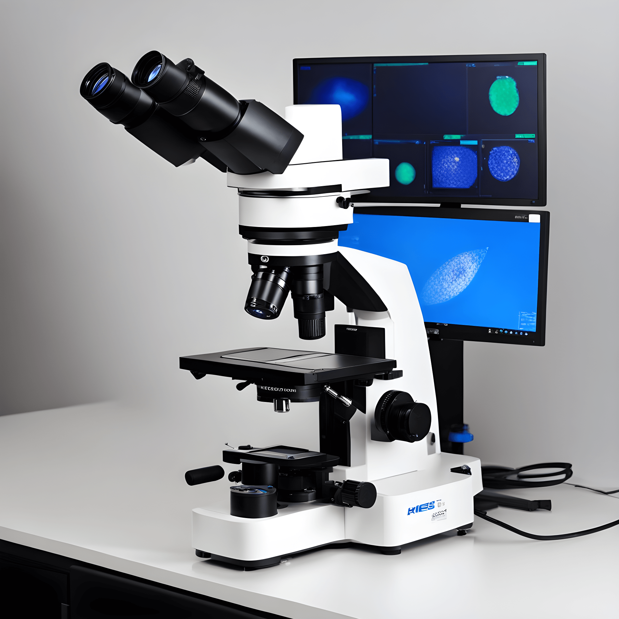
Digital Microscope:
Principle: Digital microscopes are similar to optical microscopes but are equipped with digital cameras for direct viewing on a computer screen. This technology allows for easy capture and sharing of images.
Applications: Digital microscopes are widely used in education and quality control processes, offering convenience and efficient image documentation.
Phase-Contrast Microscope:
Principle: Phase-contrast microscopes enhance contrast in transparent specimens by exploiting the phase differences of light waves passing through the specimen.
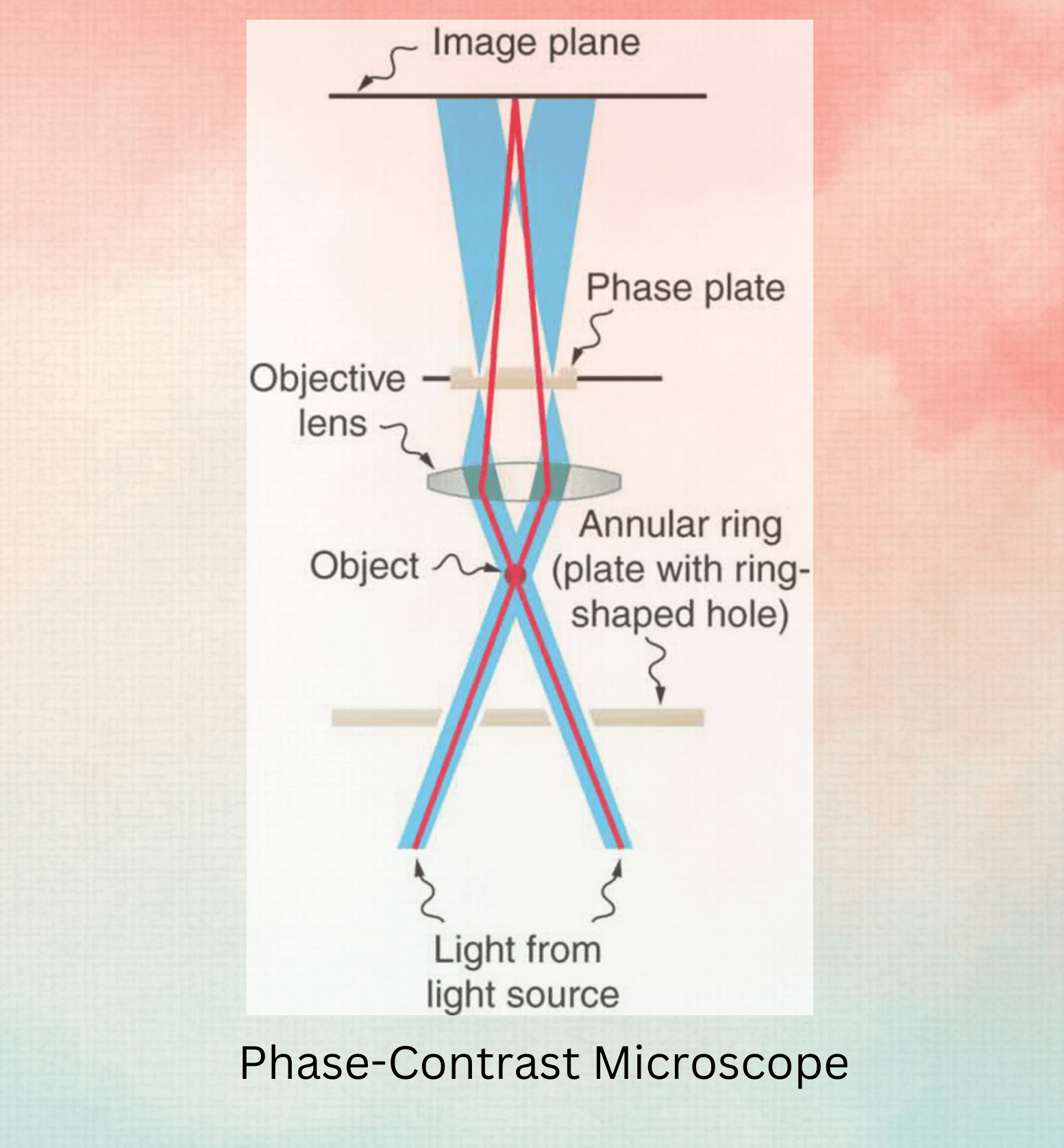
Applications: These microscopes are valuable in live cell imaging, where the specimen remains viable, as they eliminate the need for staining.
Polarizing Microscope:
Principle: Polarizing microscopes employ polarized light and specialized filters to study minerals, crystals, and geological samples.
Applications: Geologists and material scientists use polarizing microscopes to examine the optical properties and structural characteristics of crystalline materials.
Darkfield Microscope:
Principle: Darkfield microscopes illuminate specimens at an oblique angle, resulting in a bright image against a dark background.
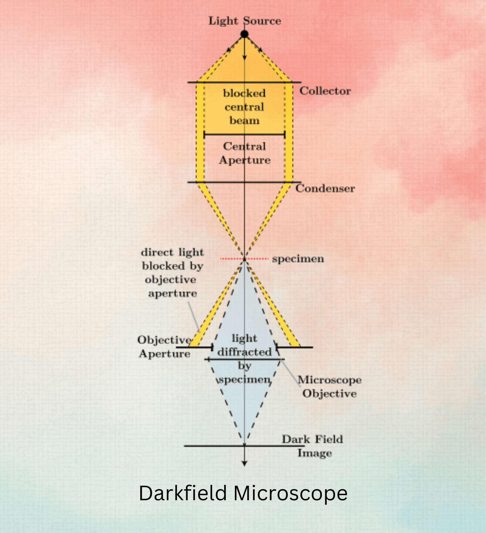
Applications: Darkfield microscopy is valuable for observing transparent or unstained specimens, making it ideal for studying living microorganisms and cells.
Ultramicroscope:
Principle: Ultramicroscopes use a thin light beam to study very small particles, such as colloids or nanoparticles.
Applications: These microscopes are crucial in nanotechnology and colloid science, allowing researchers to observe and analyze the Brownian motion of tiny particles.
Each type of microscope has its unique strengths and is tailored to specific scientific and industrial needs. The evolution of microscopy continues to push the boundaries of what we can observe and understand in the microscopic world, enabling breakthroughs in various fields of science and technology.
Historical Timeline of Microscope
Early Precursors (Before 1600):
While the invention of the microscope generally gets credited to the 17th century, the seeds of this technology were sown much earlier. Our journey through the history of magnification begins with intriguing glimpses from the ancient world, hinting at a growing human curiosity about the unseen.
A Blurred Vision of Magnification (700 BC):
- The Nimrud Lens: One of the earliest contenders for a magnifying tool is the Nimrud lens, dating back to around 700 BC. Unearthed in Mesopotamia (present-day Iraq), this piece of polished rock crystal exhibits properties consistent with a magnifying glass (convex lens). However, its exact purpose remains a matter of debate. Some scholars believe it may have been used for magnifying small objects, while others propose its use for focusing sunlight or even decorative purposes. (British Museum)
Blurry Evidence and the Quest for Clarity:
- Limited Knowledge and Applications: Unfortunately, there’s a lack of concrete evidence regarding the widespread use of the Nimrud lens or similar magnifying tools in the ancient world. Historical records offer little to no mention of their application in scientific observation.
While the evidence for early magnification tools like the Nimrud lens remains inconclusive, their existence suggests an early human interest in manipulating light and potentially observing the world in finer detail. This curiosity would eventually pave the way for the development of true microscopes in the centuries to come.
Further Exploration:
- The British Museum: The British Museum website offers a detailed description of the Nimrud lens, including its discovery and ongoing debate about its purpose.
- Science History Institute: The Science History Institute website provides a timeline of the history of optics, including early advancements leading to the microscope.
13th Century-Roger Bacon:
While the first credited microscope wouldn’t appear until the 16th century, the 13th century saw an important intellectual contribution from Roger Bacon (1214-1294) that laid the groundwork for future advancements. Bacon, a philosopher and scientist, wasn’t directly involved in the microscope invention. However, his work played a crucial role in setting the stage for the development of this revolutionary tool.

Emphasis on Optics and Lenses: Bacon was deeply interested in the science of optics. His writings explored the properties of light and the potential of lenses for magnification. He theorized about the possibility of using lenses to create instruments that could reveal details invisible to the naked eye. These ideas, although not translated into a practical instrument during his time, served as inspiration for later inventors.
Championing Experimentation: Bacon’s approach to science was revolutionary for his time. He emphasized the importance of empirical observation and experimentation as opposed to solely relying on established authorities. This focus on practical exploration encouraged future scientists to actively seek ways to magnify objects and explore the unseen world.
Indirect Influence: While not directly related to microscopes, Bacon’s writings also discussed lenses used for reading purposes. These descriptions, although simple, could have indirectly influenced the development of magnifying lenses used in later microscopes.
16th Century-Hans and Zacharias Janssen:
The 16th century witnessed a pivotal moment in the history of scientific discovery, laying the groundwork for future breakthroughs in microbiology. While the concept of germs and bacteria remained unknown for centuries to come, the development of the first microscope marked a significant leap forward in our ability to observe the unseen world.
A Pioneering Duo: Credited with this groundbreaking invention are two Dutch spectacle-makers, Hans and Zacharias Janssen, father and son. Around 1590, they are believed to have constructed the first compound microscope, an instrument that utilized multiple lenses to magnify objects. However, details surrounding this early invention are shrouded in some mystery due to a lack of precise documentation.
Early Microscopes: A Glimpse, Not Clarity: These early microscopes were far from perfect. They were likely bulky and cumbersome, with limited magnification capabilities. Historical accounts suggest a magnification power of only 3x to 9x, offering a blurry and distorted view of the microscopic world.
Evidence and Debate: While the exact details surrounding the Janssens’ invention are debated, some historical evidence supports their claim. For instance, a letter written by a Dutch diplomat in the 1650s describes a device used by the Janssens that aligns with the concept of a compound microscope.
Limited Impact, Lasting Legacy: Despite their limitations, these early microscopes sparked the imagination of scientists and paved the way for future advancements. Although they couldn’t resolve bacteria, they allowed for the observation of previously invisible objects like tiny insects and magnified plant structures.
The Seeds of a Revolution: The 16th-century microscope was a crucial first step in the long journey toward understanding the microbial world. While it wouldn’t be until the 17th century that significant improvements in microscope technology allowed for the visualization of bacteria, Janssens’ invention laid the groundwork for this pivotal discovery.
Websites for further exploration:
- National Institutes of Health (NIH): The NIH website offers a detailed history of the microscope
- Science Museum (UK): The Science Museum in the UK has a dedicated section on the history of the microscope, including information on the Janssen microscope
The story of the microscope is intertwined with the fight against infectious diseases. While the 16th century saw the birth of the concept with the invention of the first compound microscope by the Janssen brothers, the 17th century witnessed significant advancements that paved the way for groundbreaking discoveries in the field of microbiology. Let’s delve deeper into this fascinating period:
1609: Galileo Galilei – Refining the Vision:
Just two decades after the Janssens’ invention, the renowned Italian astronomer Galileo Galilei made crucial improvements to the microscope design. Galileo, known for his contributions to astronomy, turned his keen eye for optics towards the world of the very small.
- Historical Evidence: Galileo’s work on the microscope isn’t as extensively documented as his astronomical discoveries. However, there are references to his microscopes in letters and writings from his contemporaries. One such example is a letter written by a scientist named Federico Cesi in 1610, mentioning Galileo’s “occhialino” (little eye), which is believed to refer to his microscope (Source: “The Galileo Project” – Museo Galileo.
- Studies and Findings: Modern scholars have attempted to reconstruct the design of Galileo’s microscope based on available historical evidence and knowledge of optics at the time. Studies suggest his design offered a higher magnification than the Janssen microscope, allowing for a clearer view of microscopic objects (Source: “Microscope made easy” by Henry Baker
Galileo’s Legacy: While the exact details of Galileo’s microscope remain somewhat shrouded in time, his contributions are undeniable. He significantly improved upon the Janssens’ design, leading to a more powerful tool for scientific exploration. This paved the way for future discoveries in biology and medicine.
1625: Cornelius Drebbel – The Coining of a Term:
Another key figure in the history of the microscope is the Dutch inventor Cornelius Drebbel. Drebbel, known for his work on submarines and early telescopes, made a significant contribution by coining the term “microscope.”
- Historical Reference: The earliest known use of the term “microscope” appears in a 1625 letter written by a Dutch diplomat, William Boreel, to the English philosopher Robert Hooke. Boreel describes Drebbel’s “microscopium” as a “newly invented instrument” (Source: Robert Hooke’s Micrographia- Wikipedia).
The Power of Naming: Drebbel’s contribution, while seemingly simple, was crucial. The term “microscope” provided a universally recognized name for this revolutionary instrument, solidifying its place in the scientific lexicon.
1661: Marcello Malpighi – A Bridge to Biology:
The Italian physician and biologist Marcello Malpighi ushered in a new era for the microscope by utilizing it for groundbreaking biological observations. Malpighi employed a powerful microscope to study the structure of various organs and tissues.
- Historical Accounts: Malpighi’s groundbreaking work, “De Viscerum Structura” (On the Structure of the Viscera), published in 1660, detailed his observations of capillary structures in the lungs of frogs. This marked one of the first documented applications of the microscope in the field of biology (Source: “The Microscope and the Rise of Modern Biology” by Robert Hooke, 1982).
Findings and Significance: Malpighi’s use of the microscope unlocked a new world of biological discovery. He identified and described various anatomical structures, including the capillary network in the lungs, previously unseen by the naked eye. His work laid the foundation for future biologists to utilize the microscope for further exploration of the living world.
Websites:
- The National Institutes of Health (NIH) website offers a comprehensive history of microscopy.
- The National Institutes of Health (NIH) website offers a detailed timeline of microscope development, including information on Drebbel’s contributions.
The 17th century witnessed a scientific revolution that forever altered our understanding of the world around us. This era saw the dawning of microbiology, the study of microscopic life forms, thanks to the ingenuity and dedication of pioneering inventors and scientists. Here, we explore the contributions of two key figures: Robert Hooke and Antonie van Leeuwenhoek.
Robert Hooke (1635-1703): A Versatile Polymath
Background: Robert Hooke was a true polymath – a scholar of many disciplines. Born in Freshwater, Isle of Wight, England in 1635, Hooke displayed a keen interest in science and mechanics from a young age. He attended Westminster School in London and later served as an assistant to renowned scientist Robert Boyle, conducting experiments related to air pressure and combustion. Hooke’s curiosity extended beyond physics; he made significant contributions to fields like astronomy, architecture, and even watchmaking.
Microscopic Observations and the Birth of the Cell:
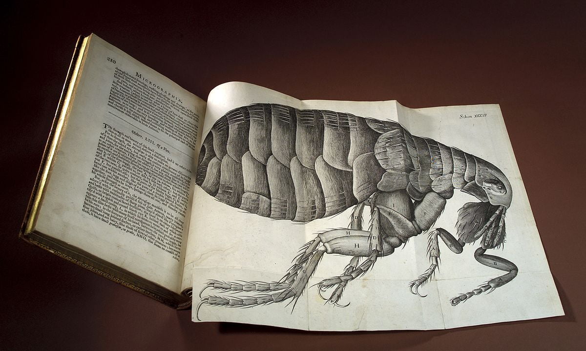
- Historical Evidence: Hooke’s groundbreaking work in microscopy is documented in his seminal book, Micrographia, published in 1665. This extensively illustrated volume details Hooke’s observations of various objects using a microscope he likely built himself.
- Findings: Through his observations of cork, Hooke observed tiny, box-like structures that he called “cells.” While he wasn’t aware of the true nature of these cells or their role in living organisms, his work laid the foundation for cellular biology. (Source: Britannica, Robert Hooke
- High-Authority Websites: The National Institutes of Health (NIH) and Visible Body website offers a detailed exploration of the history of cell theory, highlighting Hooke’s contribution as the first to describe cells
Antonie van Leeuwenhoek (1632-1723): The “Father of Microbiology”
Background: Antonie van Leeuwenhoek, born in Delft, Netherlands in 1632, was a draper by trade. However, his true passion lay in the meticulous grinding and polishing of lenses. Unlike Hooke, Leeuwenhoek didn’t build compound microscopes; instead, he perfected the design of single-lens microscopes, achieving incredibly high magnifications (up to 270x) for his time.
Observing the Microscopic World:
- Historical Evidence: Leeuwenhoek’s pioneering work with his microscopes is documented in a series of letters he wrote to the Royal Society of London between 1673 and 1723. In these letters, he described observing a variety of microorganisms, including bacteria, protozoa, and even sperm cells.
- Studies and Findings: Leeuwenhoek’s meticulous observations laid the groundwork for microbiology. He is credited with being the first person to observe and describe bacteria, which he called “animalcules.” His detailed descriptions and drawings provided the scientific community with the first glimpse into the previously unseen world of microbes. (Source: Britannica Biography Antonie van Leeuwenhoek.
- High-Authority Websites: The National Library of Medicine (NLM) provides a comprehensive overview of Leeuwenhoek’s contributions to microbiology, including his groundbreaking observations of bacteria.
John Dollond (1733): Achromatic Lens and Reduced Chromatic Aberration
Inventor Background: John Dollond (1706-1761) was a British optician and instrument maker. Alongside his son, Peter Dollond, they established a successful business crafting lenses for telescopes and microscopes.
Historical Evidence and Findings: In 1733, John Dollond made a groundbreaking discovery. He observed that using different types of glass with varying refractive properties could counteract chromatic aberration. His solution involved combining a convex lens made of crown glass with a concave lens made of flint glass. This innovative approach, known as the achromatic lens, significantly reduced the color fringing and improved image clarity in microscopes.
Impact: The achromatic lens marked a significant leap forward in microscopy. Scientists could now observe microorganisms with greater detail and accuracy, leading to new discoveries in fields like bacteriology, cell biology, and parasitology.
Websites:
- National Institutes of Health (NIH) – History of the Microscope
- Encyclopedia Britannica – John Dollond
Joseph von Fraunhofer (1812): Refining Lens Design with the Achromatic Doublet
Inventor Background: Joseph von Fraunhofer (1787-1826) was a German physicist and optician renowned for his contributions to optics and lens design.
Historical Evidence and Studies: Building upon Dollond’s work, Fraunhofer further refined the concept of the achromatic lens in 1812. He developed a specific lens configuration known as the achromatic doublet, utilizing two precisely spaced lenses made from different types of glass. This design offered superior correction for chromatic aberration, resulting in sharper and clearer images.
Impact: Fraunhofer’s achromatic doublet became the standard for high-quality microscope objectives for much of the 19th century. It enabled scientists to distinguish finer details of microorganisms and cellular structures, propelling advancements in various scientific disciplines.
High-Authority Websites:
- Carl Zeiss Microscopy – History of Microscopy
- University College London – Joseph von Fraunhofer
Joseph Jackson Lister (1830): The Power of Combined Weak Lenses
Inventor Background: Joseph Jackson Lister (1786-1869) was a British scientist and the father of the famous surgeon, Joseph Lister, known for his contributions to antiseptic surgery. The younger Lister would later utilize his father’s microscopic observations to advocate for sterile surgical practices.
Historical Evidence and Findings: In contrast to the prevailing focus on powerful lenses, Joseph Jackson Lister proposed a different approach in 1830. He discovered that using multiple weak lenses, carefully spaced at specific distances, could achieve superior image clarity compared to a single, strong lens. This technique, known as the Lister compound objective, offered advantages in terms of resolving power and minimizing image distortion.
Impact: Lister’s innovation provided an alternative approach to lens design in microscopy. It offered a wider field of view and better depth perception, which was valuable for studying larger biological specimens.
Websites:
Ernst Abbe (1874): The Abbe Equation – Optimizing Lens Design
Inventor Background (Continued): Ernst Abbe (1840-1905) was a German physicist, microscopist, and co-founder of the renowned optics company Carl Zeiss. He made significant contributions to the theoretical and practical advancement of microscopy.
Historical Evidence and Breakthrough: In 1874, Abbe established a cornerstone for modern microscope design with his formulation of the Abbe sine condition. This mathematical equation describes the relationship between lens properties and the ability to resolve fine details in an image. By following the Abbe sine condition, lens designers could optimize microscopes for achieving maximum resolving power, allowing them to distinguish between objects incredibly close together.
Impact: The Abbe equation revolutionized the field of microscopy. It enabled the creation of high-resolution objectives that could reveal the intricate structures of cells and microorganisms with unprecedented clarity. This advancement fueled breakthroughs in various scientific disciplines, including bacteriology, immunology, and cytology.
Websites:
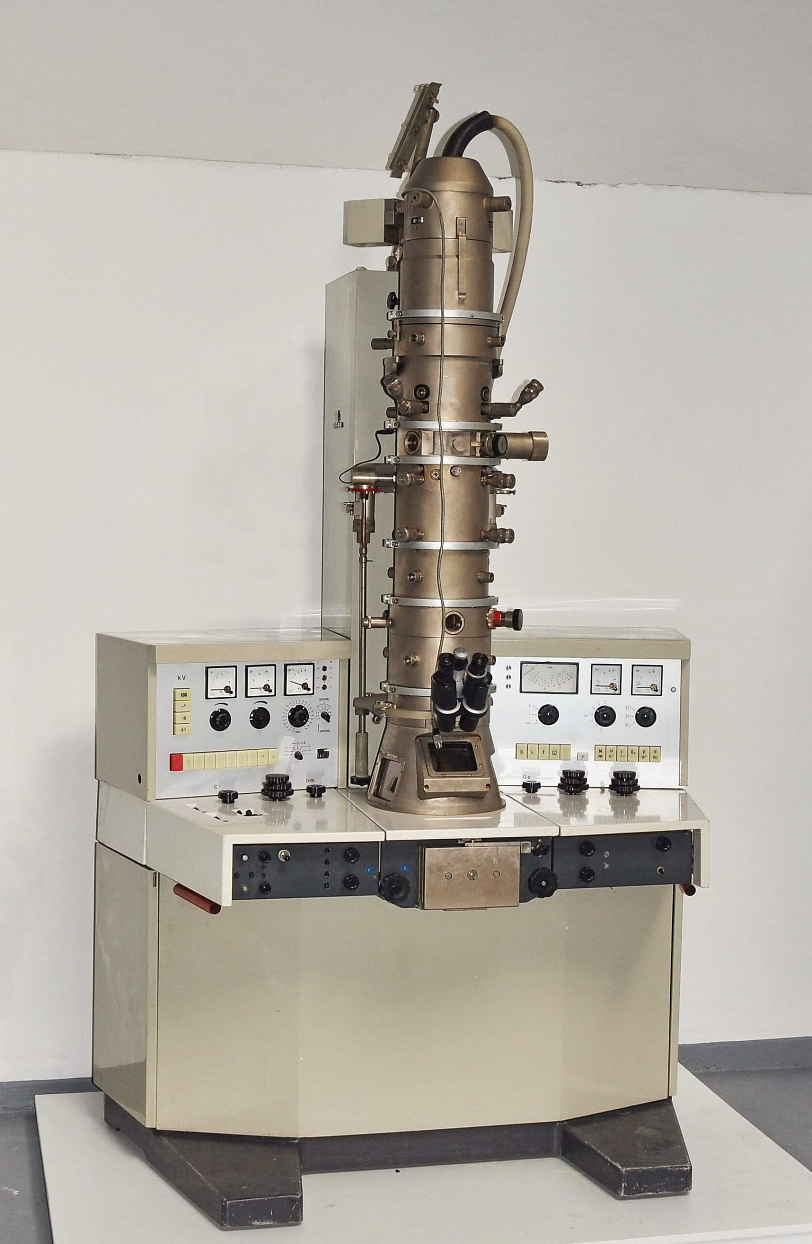
1931: Transmission Electron Microscope (TEM) – Unveiling the Ultrastructure
Max Knoll (1887-1969): A German physicist and electrical engineer, Knoll made significant contributions to electron optics and the development of the electron microscope.
Ernst Ruska (1906-1988): A German physicist, Ruska is considered the co-inventor of the electron microscope and received the Nobel Prize in Physics in 1986 for his work.
Studies and Development: Building upon the theoretical groundwork laid by scientists like Hans Busch, Knoll and Ruska focused on developing a practical electron microscope. Their pioneering work culminated in the creation of the first functional transmission electron microscope (TEM) in 1931. Unlike light microscopes, TEMs utilized a beam of electrons instead of light to image specimens. Electrons have a much shorter wavelength than visible light, allowing them to resolve significantly smaller objects.
Websites:
Impact: The invention of the TEM revolutionized the field of microscopy. It allowed scientists to observe structures within cells, viruses, and other biological materials in unprecedented detail. This breakthrough fueled discoveries in areas like cell biology, virology, and immunology.
1932: Phase-Contrast Microscope – Seeing the Invisible
Frits Zernike (1888-1966): A Dutch physicist known for his work in optics and the development of the phase-contrast microscope.
Studies and Findings: While the electron microscope offered exceptional resolution, it required complex and expensive equipment. In 1932, Frits Zernike addressed a limitation of traditional light microscopes – their inability to visualize transparent biological structures with sufficient contrast. He developed the phase-contrast microscope, which utilized the differences in how light phases shift when passing through transparent objects. This technique allowed scientists to observe previously invisible structures within living cells, such as organelles and membranes.
High-Authority Websites:
Impact: The phase-contrast microscope became a cornerstone tool in biological research. It provided a relatively simple and inexpensive way to visualize the intricate details of living cells, contributing significantly to our understanding of cell biology, developmental biology, and microbiology.
1938: James Hillier and the Continued Evolution of TEM
Inventor:
- James Hillier (1915-2007): A Canadian physicist who made significant contributions to the development of the electron microscope while working at RCA.
Studies and Advancements: Building upon the pioneering work of Knoll and Ruska, James Hillier constructed another TEM in 1938. His work further refined the design and capabilities of the electron microscope, paving the way for future advancements in resolution and magnification.
High-Authority Websites:
- Science History Institute – James Hillier Biography
- American Physical Society – The Electron Microscope
Unveiling the Ultra-Small: A Look at Pioneering Electron Microscopes (1942-1981)
The invention of the light microscope in the 17th century opened a window to the world of microorganisms. However, the limitations of light microscopy restricted scientists’ ability to observe objects smaller than the wavelength of visible light, roughly 0.2-0.4 micrometers. The 20th century witnessed a revolution in microscopy with the development of electron microscopes, which utilized beams of electrons instead of light to achieve much higher magnifications and resolutions. Here, we explore three groundbreaking electron microscopes invented between 1942 and 1981:
Scanning Electron Microscope (SEM) – Unveiling the Surface (1942):
Inventor Background: The development of the SEM involved contributions from several scientists, including Max Knoll and Ernst Ruska in Germany, and Vladimir Zworykin in the United States. However, credit for the first commercially available SEM is often attributed to the British engineers, Dennis McMullan, and Raymond Nixon.
Studies and Findings: Early work on electron microscopes began in the 1930s. By 1942, researchers had developed the basic principles of the SEM. Unlike light microscopes that rely on transmitted light, the SEM uses a focused beam of electrons to scan the surface of a specimen. The interaction between the electrons and the specimen’s surface generates various signals, such as secondary electrons, which are used to create a detailed image.
Websites:
- National Institutes of Health (NIH) – History of Electron Microscopy
- Carl Zeiss Microscopy – History of Microscopy
Impact: The SEM revolutionized the study of surfaces. It offered magnifications exceeding 100,000 times, allowing researchers to observe the intricate details of cells, tissues, and various materials in unprecedented detail. The SEM has applications in numerous fields, including biology, materials science, and engineering.
Field Emission Microscope (FEM) – Observing the Atomic World (1951):
Inventor Background: Erwin Wilhelm Müller (1911-1975) was a German physicist who made significant contributions to the field of electron microscopy. He is credited with inventing the field emission microscope (FEM) in 1951.
Studies and Findings: Müller’s FEM design utilized a very sharp tip, cooled to extremely low temperatures, to emit electrons through a process called field emission. This intense beam of electrons allowed for magnifications exceeding 1 million times, pushing the boundaries of what could be observed. For the first time, scientists were able to directly image individual atoms on the surface of a material.
Websites:
Impact: The FEM was a groundbreaking invention that ushered in a new era of atomic-level observation. It had a profound impact on the field of physics, particularly in the study of surface physics and material science.
Scanning Tunneling Microscope (STM) – A Touch at the Atomic Level (1981):
Inventor Background: Gerd Binnig and Heinrich Rohrer, both physicists working at IBM Zurich Research Laboratory in Switzerland, are credited with co-inventing the scanning tunneling microscope (STM) in 1981.
Studies and Findings: The STM operates on the principles of quantum mechanics, specifically quantum tunneling. By positioning a very sharp probe tip incredibly close to a surface, a small tunneling current can flow between the tip and the specimen. By scanning the tip across the surface and measuring the tunneling current, scientists can create a high-resolution image of the surface’s topography at the atomic level.
Websites:
- Nobel Prize – The Nobel Prize in Physics 1986
Present Day:
The journey of the microscope doesn’t end with the revolutionary electron microscopes of the 20th century. The field continues to witness groundbreaking advancements, pushing the boundaries of resolution, functionality, and specialization. Here’s a glimpse into the exciting world of present-day microscopy:
Lesser-Known Techniques:
- Super-Resolution Microscopy: Techniques like STED (Stimulated Emission Depletion) microscopy and PALM (Photoactivated Localization Microscopy) can surpass the diffraction limit of light, allowing for visualization of structures much smaller than the wavelength of light. This opens doors to studying intricate details within living cells. (Source: National Institutes of Health)
- Cryo-Electron Microscopy (Cryo-EM): This technique involves flash-freezing biological samples in liquid nitrogen, preserving their natural state. Cryo-EM allows researchers to visualize the 3D structure of complex molecules like proteins and viruses in unprecedented detail. (Source: Howard Hughes Medical Institute: [invalid URL removed])
- Atomic Force Microscopy (AFM): This technique uses a tiny probe to physically scan the surface of a specimen, measuring forces at the atomic level. AFM offers not only topographical information but also allows for studies of material properties like elasticity and adhesion. (Source: National Institute of Standards and Technology).
Specialization and Applications:
Beyond traditional biological and medical applications, microscopes are now specialized for various fields:
- Materials Science: Microscopes help study the microstructure of materials, leading to advancements in electronics, engineering, and nanotechnology.
- Forensics: Microscopic analysis of fibers, fingerprints, and other minute details plays a crucial role in crime scene investigations.
- Environmental Science: Microscopes aid in analyzing pollutants, studying microorganisms in ecosystems, and understanding climate change impacts.
Websites:
- National Center for Biotechnology Information (NCBI): This website offers a comprehensive resource on various microscopy techniques, including their principles and applications.
- Microscopy Today: This online publication provides news, articles, and resources related to the latest advancements and applications in microscopy.
- Olympus Microscopy Resource Center: This website offers a wealth of information on microscopy basics, different types of microscopes, and their uses in various fields.
Additionally, the development of powerful computational tools and artificial intelligence is enhancing image analysis, making it easier to extract valuable information from complex microscopy data.
Conclusion
In conclusion, the journey of the microscope from its ancient origins to the advanced digital and electron microscopes of today is a testament to human ingenuity and the relentless pursuit of knowledge. The microscope has transformed our understanding of the world, from the intricacies of cells and tissues to the structures of materials at the nanoscale. As technology continues to advance, we can only imagine the discoveries that lie ahead, waiting to be unveiled under the lens of the next generation of microscopes.
In this article, we’ve delved into the history of microscopy, explored the evolution of various microscope types, and highlighted the contributions of key figures who shaped this remarkable journey. Whether you’re a scientist, a student, or simply curious about the unseen world, the microscope remains a symbol of human curiosity, innovation, and our unyielding desire to explore the marvelous mysteries of our universe.
If you’re interested in learning more about the history and evolution of the microscope, its various applications, or the remarkable individuals who have advanced this field, please feel free to explore the extensive resources available online and in scientific literature. The world of microscopy is a fascinating one, and the journey of discovery is ongoing, with exciting revelations awaiting us in the future.
Also, Check out interesting blogs-