Introduction: Diagnostic Imaging History
Diagnostic imaging has revolutionized healthcare by offering a window into the human body without invasive procedures. From identifying fractures with X-rays to detecting tumors with MRIs, the field has transformed medicine, saving countless lives and enhancing our understanding of diseases. The diagnostic imaging history is a story of relentless innovation, starting from rudimentary tools and evolving into cutting-edge technologies like CT scans vs MRIs, each offering unique advantages in advanced diagnostics.
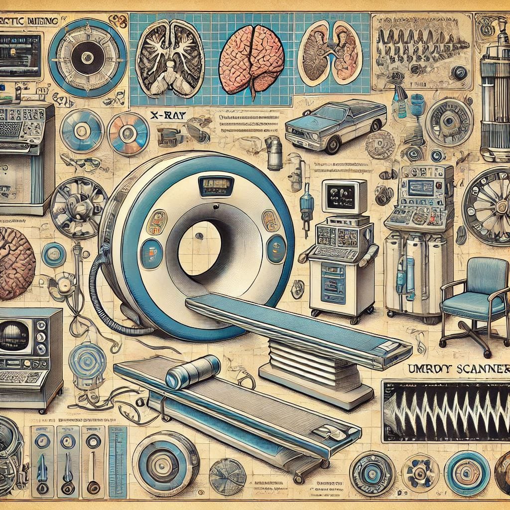
The journey of imaging reflects the synergy of science and medicine, driven by the need for precision and early intervention. Beyond its clinical role, medical imaging innovation has enabled researchers to explore human anatomy and physiology like never before. As we look to the future of medical imaging, the integration of AI, portable devices, and sustainable practices promises to make diagnostics more efficient, accessible, and eco-friendly—a leap toward sustainable healthcare.
The Origins of Diagnostic Imaging
Discovery of X-rays by Wilhelm Röntgen (1895)
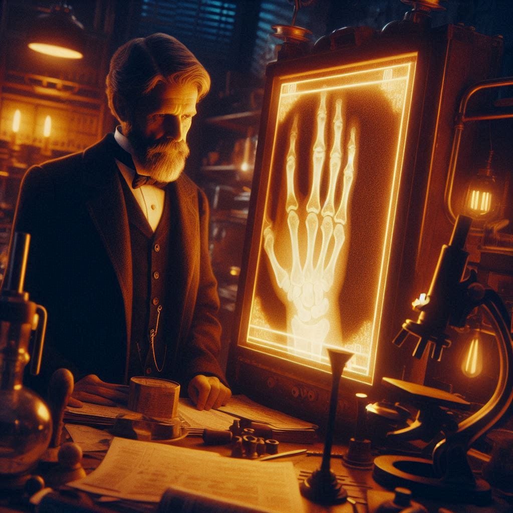
The origins of diagnostic imaging history are rooted in the groundbreaking discovery of X-rays by Wilhelm Conrad Röntgen on November 8, 1895. While experimenting with cathode ray tubes in a darkened lab, Röntgen noticed a fluorescent screen glowing unexpectedly. This phenomenon, caused by an unknown form of invisible radiation, prompted him to conduct further studies. He captured the first X-ray image of his wife’s hand, where her bones and wedding ring were vividly visible. This image became a defining moment in the evolution of X-rays, showcasing their potential for non-invasive medical diagnostics. For his contributions, Röntgen was awarded the inaugural Nobel Prize in Physics in 1901, cementing the significance of his discovery.

Early Applications and Immediate Impact
Within months of Röntgen’s discovery, X-rays were applied in various medical and industrial contexts. Physicians used them to locate fractures, embedded objects like bullets, and dental issues. Early devices were primitive, often exposing operators and patients to significant radiation without safeguards. Despite these challenges, the discovery rapidly gained traction, marking a new era in the history of radiology and transforming medical practice.
Technological Advancements
The development of radiographic equipment advanced rapidly after the initial discovery. In 1896, Thomas Edison introduced fluoroscopy, allowing live imaging of internal structures. Innovations like the introduction of contrast agents improved the visualization of soft tissues, while the invention of rotating anode tubes significantly enhanced image resolution. These technological strides transitioned X-rays from rudimentary tools to sophisticated instruments integral to healthcare. The advent of protective measures, such as lead shielding and film-based detectors, addressed early concerns about radiation exposure.
Challenges and Safety Concerns
The journey of radiographic technology wasn’t without hurdles. In the early 20th century, practitioners and patients often suffered radiation burns due to prolonged exposure. The lack of understanding about the long-term effects of X-rays posed significant health risks. Safety standards gradually emerged, driven by research on radiation’s biological impact. By the mid-1900s, protective gear and regulated protocols became mandatory, significantly reducing risks associated with X-ray imaging.
Transition to a Specialized Field
As X-ray technology matured, the field of radiology emerged, integrating diagnostic imaging with therapeutic applications. By the early 20th century, radiologists specialized in the interpretation of X-rays, and the field expanded to include subspecialties like interventional radiology and oncology imaging. This transition highlighted the broader impact of diagnostic imaging on healthcare and its role in shaping modern medical practices.
Fresh Insights:
- Pre-Röntgen imaging practices included crude methods like transillumination using candles or rudimentary light sources. These techniques lacked precision, underscoring the revolutionary nature of X-ray discovery.
- Newly discovered manuscripts reveal the use of X-rays in anthropology and archaeology as early as the 1900s, aiding the examination of mummies and ancient artifacts.
- Uncommon applications, such as X-rays in early aviation (e.g., inspecting plane parts), illustrate the broader impact of Röntgen’s invention beyond healthcare.
The birth of diagnostic imaging history through Röntgen’s discovery of X-rays marked the dawn of a technological revolution, fundamentally altering medicine and offering unparalleled insights into the human body. This journey from rudimentary cathode experiments to the establishment of radiographic technology laid the foundation for modern imaging systems like CT scans and MRIs, which continue to save lives globally.
The Evolution of Imaging Modalities
The evolution of X-rays marked the beginning of a remarkable journey in diagnostic imaging history, setting the stage for the development of advanced imaging techniques that transformed medicine.
The Advent of CT Scans (1970s): A New Dimension in Imaging
The 1970s saw the introduction of CT scans (Computed Tomography), a technology that revolutionized trauma care and diagnostic precision. CT scans provided detailed cross-sectional images of the body by combining multiple X-ray images with computer processing.
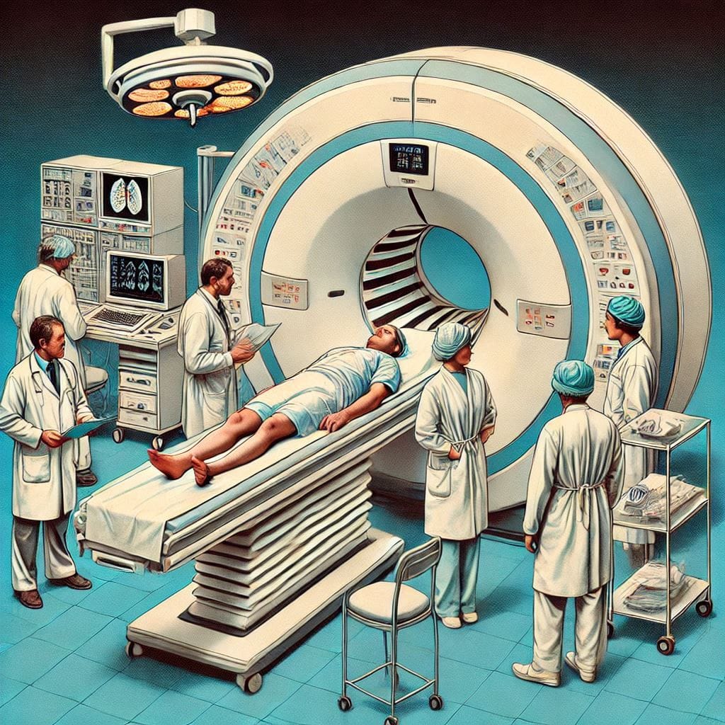
This innovation offered the ability to visualize internal organs and tissues in 3D, addressing challenges that plain X-rays couldn’t overcome, such as differentiating between soft tissues and detecting internal bleeding. The rapid imaging capability of CT scans made them invaluable in emergency settings, particularly for head injuries and strokes. This advancement cemented its place in diagnostic imaging history, offering clarity and speed in life-saving scenarios.
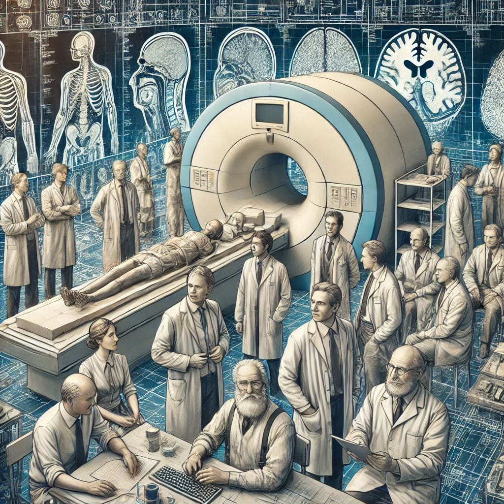
The Rise of MRI Technology (1980s): Soft Tissue Imaging Reimagined
The 1980s introduced MRI technology (Magnetic Resonance Imaging), a non-invasive method that transformed the imaging of soft tissues such as the brain, muscles, and ligaments. Unlike X-rays and CT scans, MRI does not use ionizing radiation but relies on magnetic fields and radio waves. This technology is particularly suited for identifying abnormalities in the central nervous system, such as brain tumors or spinal cord injuries. MRI’s ability to produce highly detailed images without harming tissues was a significant leap in imaging techniques, expanding diagnostic capabilities while ensuring patient safety.
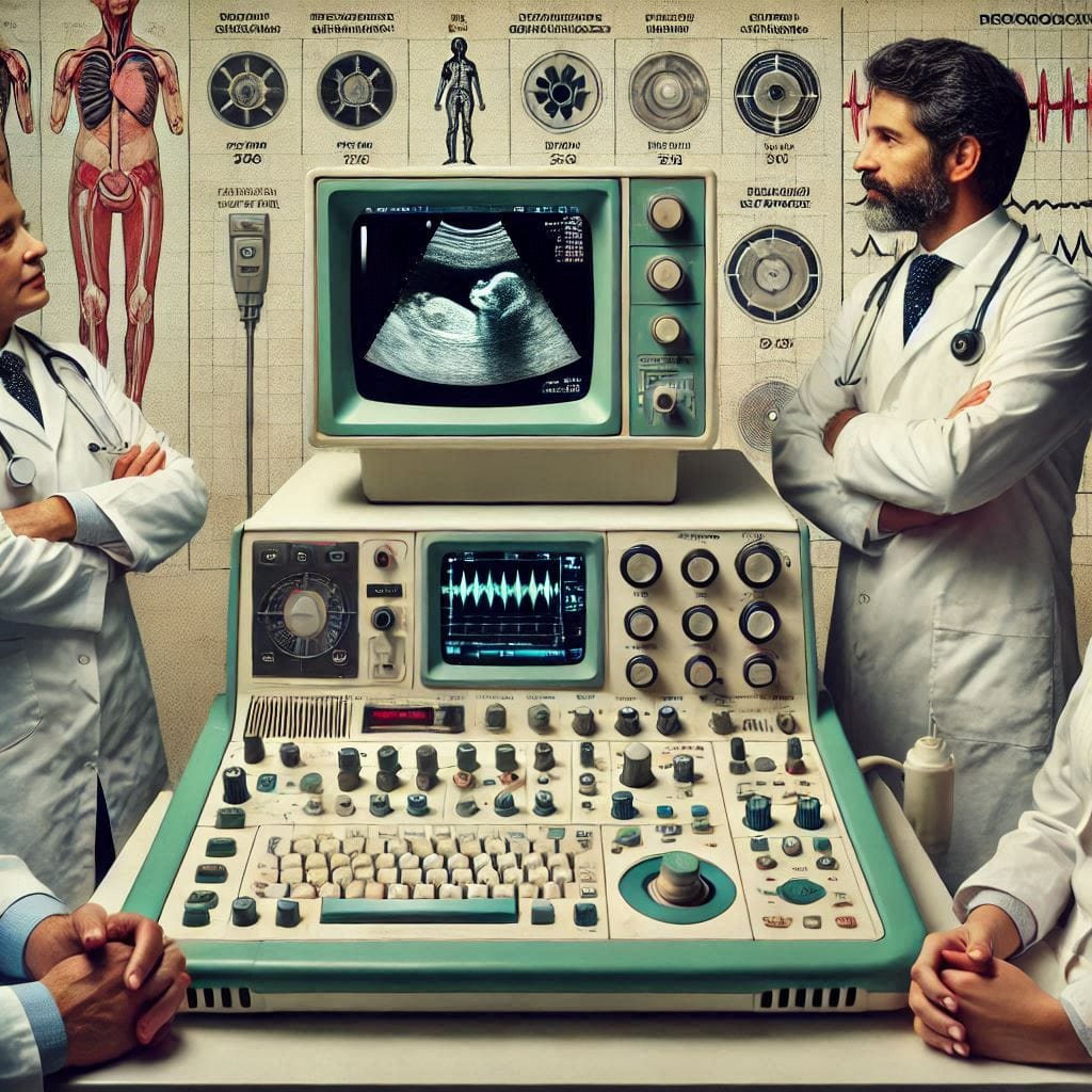
The Evolution of Ultrasound: A Safe and Versatile Tool
The origins of ultrasound date back to the mid-20th century, but its rapid advancement made it a cornerstone of diagnostic imaging history by the 1980s and 1990s. Known for its safety and accessibility, ultrasound uses sound waves to generate images, making it indispensable for prenatal care, cardiovascular diagnostics, and musculoskeletal imaging. Unlike MRI and CT scans, ultrasound does not use radiation, making it a preferred choice for vulnerable populations, such as pregnant women and pediatric patients. The ultrasound evolution has also led to portable and handheld devices, expanding access to imaging in remote and resource-limited settings.
Fresh Insights:
- CT scans and beyond: Recent research highlights ongoing innovations in CT technology, such as dual-energy CT, which improves differentiation of tissue types and enhances diagnostic accuracy.
- MRI advancements: Functional MRI (fMRI) has expanded the scope of MRI to include mapping brain activity, particularly for pre-surgical planning and research into neurological disorders.
- Ultrasound innovation: The integration of AI in ultrasound imaging has improved diagnostic accuracy and reduced operator dependency, a significant leap in its evolution.
The evolution of X-rays catalyzed the development of diverse imaging techniques, each tailored to specific medical needs. From the 3D capabilities of CT scans to the unparalleled clarity of MRI technology and the safety of ultrasound evolution, these modalities collectively transformed how we diagnose and treat diseases. This continuous innovation underscores the profound impact of diagnostic imaging in advancing global healthcare.
CT Scan vs MRI: A Detailed Comparison
The debate between CT scan vs MRI is integral to understanding modern imaging techniques. These diagnostic tools cater to unique medical needs, with distinctions rooted in their underlying technology, applications, and practicality.
Technological Foundations
- CT Scan: Utilizes X-rays combined with computer processing to generate cross-sectional images. Its strength lies in capturing detailed images of bones, blood vessels, and internal injuries in emergencies.
- MRI: Leverages magnetic fields and radio waves to produce highly detailed images, particularly of soft tissues. This makes MRI ideal for diagnosing conditions in the brain, spine, joints, and muscles.
The difference in these technologies means CT scans excel in visualizing dense structures, while MRI provides unparalleled clarity for soft tissue evaluation.
Best Applications
- CT Scan Advantages:
- Emergency trauma care: Rapid imaging is critical for detecting internal bleeding, fractures, or organ damage.
- Lung and abdominal assessments: Particularly effective for diagnosing conditions like pneumonia or appendicitis.
- MRI Benefits:
- Soft tissue evaluation: Preferred for identifying ligament tears, brain disorders, or spinal cord injuries.
- Neurological and cardiac imaging: Functional MRI (fMRI) can even map brain activity, aiding in surgical planning.
Understanding the roles of these diagnostic tools is key to their effective application in diverse medical scenarios.
Cost, Speed, and Accessibility
- Speed: CT scans typically take only a few minutes, making them invaluable in urgent situations. MRI scans, however, can take 30-60 minutes, requiring patient stillness for optimal results.
- Cost: MRI is generally more expensive than CT, due to its advanced technology and longer scan times.
- Accessibility: CT scanners are more widespread, especially in resource-limited settings, while MRI machines are typically found in well-equipped facilities.
Fresh Insights
- Recent advancements in CT scan technology include low-dose CT for minimizing radiation exposure, especially for cancer screening.
- Emerging innovations in MRI, like portable MRI machines, are improving access to this powerful diagnostic tool in underserved areas.
Both CT scans and MRIs play vital roles in modern diagnostics, each excelling in specific applications. While CT scan advantages include speed and accessibility, MRI benefits lie in its precision for soft tissue imaging. This balance ensures that these imaging techniques remain indispensable in advancing healthcare.
Both CT scans and MRIs are critical diagnostic tools, but they serve different purposes in medical imaging. Below is a comprehensive comparison:
| Feature | CT Scan | MRI |
|---|---|---|
| Technology | Utilizes X-rays and computer algorithms to create cross-sectional images. | Employs magnetic fields and radio waves to generate detailed images of soft tissues. |
| Radiation Exposure | Yes, involves ionizing radiation (lower doses in modern machines). | None, making it safer for sensitive populations (e.g., pregnant women, children). |
| Best Applications | Effective for visualizing dense structures like bones, blood vessels, and lungs. | Superior for imaging soft tissues, including brain, muscles, and spinal cord. |
| Speed | Faster (5–10 minutes), ideal for emergency trauma cases. | Slower (30–60 minutes), not suitable for emergencies. |
| Cost | More affordable (approximately $500–$1,500 per scan). | Higher cost (approximately $1,000–$5,000 per scan). |
| Availability | Widely accessible, even in rural or resource-limited areas. | Less common, particularly in underserved regions. |
| Recent Innovations | Low-dose CT scans reduce radiation; dual-energy CT improves soft tissue imaging. | Portable MRIs increase accessibility; functional MRI (fMRI) aids real-time brain mapping. |
Both imaging techniques have distinct advantages and limitations, making them complementary rather than competitive in the field of diagnostic imaging history.
Challenges in Modern Medical Imaging
Medical imaging technologies have revolutionized diagnostics and treatment planning. However, several critical challenges remain in ensuring these advancements benefit patients equitably and safely. Below is an expanded analysis of the most pressing issues in modern medical imaging.
Radiation Exposure Risks
Medical imaging modalities like CT scans and X-rays, while indispensable, expose patients to ionizing radiation. Prolonged or repeated exposure, particularly for patients undergoing routine monitoring, increases the risk of radiation-induced conditions such as cancer. Efforts to develop low-dose imaging technologies are ongoing, but progress remains slow due to technological and economic constraints.
Accessibility and Equity
Access to advanced imaging technologies like MRI and CT is highly unequal across the globe. In low-resource settings, many healthcare facilities lack the infrastructure or funding to install and maintain these devices. This creates disparities in diagnostic accuracy and health outcomes, particularly in rural and underserved regions. Bridging this gap requires significant investment in mobile imaging units and telehealth initiatives.
AI and Data Bias
The integration of artificial intelligence (AI) in imaging introduces ethical concerns, including biases in diagnostic algorithms. These biases can arise from non-representative training datasets, leading to disparities in diagnostic accuracy across different demographic groups. For instance, AI systems may underperform for populations underrepresented in the training data, potentially exacerbating health inequities.
Economic Barriers
High costs associated with acquiring, operating, and maintaining advanced imaging systems make them unaffordable for smaller hospitals and clinics. Additionally, the cost burden often extends to patients, limiting access to timely diagnostics for those without adequate insurance coverage.
Ethical Concerns in Data Handling
The growing use of AI in diagnostics brings challenges in ensuring patient data privacy and security. The ethical use of sensitive health data, particularly in regions with lax data protection laws, is an ongoing concern. Robust frameworks are needed to ensure patient consent and secure handling of medical information.
Sustainability Challenges
Medical imaging devices consume significant energy and produce electronic waste, raising environmental concerns. Developing sustainable imaging technologies and implementing recycling programs for outdated machines are essential steps toward minimizing their ecological footprint.
Addressing the Challenges
To overcome these barriers, a multi-faceted approach is essential. This includes:
- Advancing low-dose radiation technologies to reduce risks.
- Expanding global access through portable imaging systems and government subsidies.
- Ensuring AI models are diverse and unbiased by incorporating representative datasets.
- Promoting green imaging innovations to address environmental concerns.
These solutions could pave the way for equitable and sustainable healthcare while maintaining the diagnostic precision that modern imaging offers.
The Future of Medical Imaging
The future of medical imaging is being shaped by groundbreaking advancements in technology, promising to revolutionize healthcare. These innovations are driving a shift toward more accurate, accessible, and sustainable diagnostic solutions.
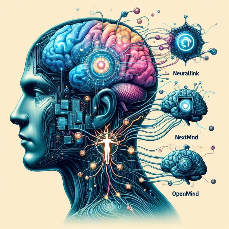
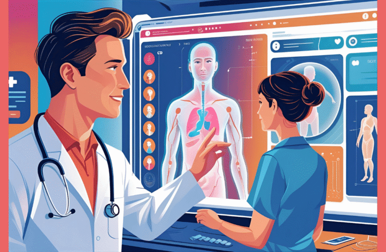
AI Integration: Real-Time Diagnostics and Predictive Analytics
The incorporation of AI in imaging is transforming diagnostics. AI algorithms analyze imaging data in real-time, detecting abnormalities with unparalleled precision. For example, AI can identify early signs of diseases such as cancer or neurological disorders that may be missed by human observers. Predictive analytics powered by machine learning can also forecast patient outcomes, allowing for personalized healthcare strategies tailored to individual needs.
Key advancements include:
- Automated detection of anomalies in radiology reports.
- Predictive modeling for treatment efficacy and patient recovery times.
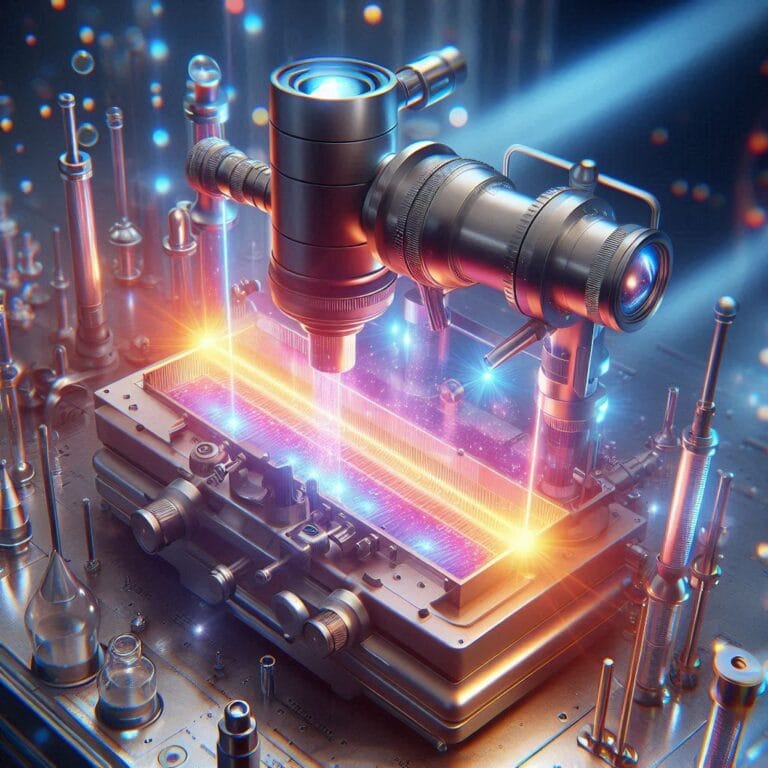
Quantum Imaging: Redefining Precision
One of the most promising frontiers in the future of medical imaging is quantum imaging. By leveraging quantum entanglement and advanced photon technologies, quantum imaging offers unparalleled clarity, especially for soft tissues and molecular diagnostics. This technology has the potential to:
- Visualize structures at the nanoscale.
- Significantly reduce radiation exposure compared to traditional methods.
- Improve the sensitivity and specificity of imaging for rare or hard-to-diagnose conditions.
While still in experimental stages, quantum imaging is poised to become a game-changer in medical imaging innovation.
Portable Imaging Devices for Point-of-Care Applications
The development of portable imaging devices is making diagnostics more accessible, especially in remote or underdeveloped regions. These compact devices provide high-quality imaging at the patient’s bedside, enabling rapid diagnosis and treatment in emergency settings. Examples include:
- Handheld ultrasound devices for cardiovascular and prenatal care.
- Mobile CT and MRI units designed for field hospitals and disaster zones.
Such innovations bridge the gap in healthcare accessibility and ensure equitable diagnostic services.
Sustainability: Energy-Efficient Machines and Waste Reduction
Modern imaging systems are often energy-intensive, contributing to high operational costs and environmental impact. Future innovations aim to:
- Develop energy-efficient imaging machines that reduce power consumption.
- Implement recycling programs for outdated equipment to minimize electronic waste.
- Adopt biodegradable components in imaging accessories to promote sustainable healthcare.
These initiatives align with global efforts to make healthcare greener and more responsible.
The future of medical imaging lies at the intersection of technological innovation and human-centric care. Advancements in AI in imaging, quantum imaging, and portable devices promise to enhance diagnostic accuracy and accessibility, while sustainability efforts ensure a reduced environmental footprint. Together, these innovations will redefine healthcare, making it more precise, inclusive, and forward-looking.
A Vision for Tomorrow: Imaging’s Role in Healthcare
As healthcare advances, medical imaging innovation will be pivotal in improving patient care. The future of healthcare relies on technologies that enhance not just diagnostic accuracy but also preventive care and personalized treatment strategies. Let’s explore the vital roles imaging will play in shaping sustainable healthcare and its integration with other emerging technologies.
Imaging’s Role in Enhancing Preventive Care
Medical imaging innovation is crucial in the early detection of diseases, often before symptoms manifest. Advances in imaging technology are enhancing the ability to screen for conditions such as cancers, cardiovascular diseases, and neurological disorders at their earliest stages. Early diagnosis allows for more effective treatment plans and a focus on preventive care. This approach can:
- Improve survival rates by identifying diseases at a curable stage.
- Decrease healthcare costs by minimizing the need for extensive treatments.
- Promote wellness-focused healthcare models.
These innovations are setting the stage for a future where personalized diagnostics can be based on regular, non-invasive imaging assessments, reducing the need for reactive medicine.
The Promise of Combining Imaging and Genetics for Individualized Treatment
One of the most groundbreaking intersections of medical imaging innovation and personalized care is the integration of imaging with genomics. By combining imaging data with genetic information, physicians can create personalized diagnostics and tailor treatment strategies specific to an individual’s genetic makeup. For example:
- Imaging can detect structural changes in organs and tissues, while genetic data can explain why these changes are occurring.
- This fusion enables more targeted interventions for diseases like cancer, where treatment can be customized based on both the location of the tumor and the genetic mutations driving its growth.
The combination of imaging technology advancements and genetic research is transforming the treatment landscape, moving us closer to individualized medicine.
The Idea of a “Digital Twin” for Real-Time Monitoring
A digital twin is a virtual replica of a patient’s body, created by merging imaging data with real-time health information. This technology can model organs and tissues in high detail, allowing physicians to monitor a patient’s health continuously. Using this model, doctors can simulate how the body will respond to different treatments and make adjustments before starting the actual intervention. The key benefits include:
- Real-time monitoring of chronic conditions like heart disease and diabetes.
- Predictive capabilities, where advanced diagnostics can forecast disease progression or recovery.
- Enhanced personalized diagnostics with the ability to simulate potential treatments based on specific patient data.
The digital twin concept could revolutionize the way healthcare professionals approach both preventative care and chronic disease management.
Conclusion
The evolution of medical imaging has revolutionized healthcare, progressing from X-rays to advanced AI-driven diagnostics. As we look ahead, sustainable healthcare and medical imaging innovation are poised to further transform patient care by integrating cutting-edge technologies like AI, genomics, and digital twins. These innovations will not only enhance the precision of diagnostic imaging but also ensure that healthcare is more personalized, preventative, and accessible.
By continuing to invest in these technologies, the future holds the promise of a healthcare system where early diagnosis and individualized treatments become the norm, improving outcomes and quality of life for all. Future diagnostic tools are shaping a new era of healthcare, providing insights into better patient management and revolutionizing the way we approach treatment and wellness.
Related Articles
- The History of Medtech
Discover the evolution of medical technologies that paved the way for modern advancements. - 3-D Printed Hearts
Learn how cutting-edge 3-D printing technology is revolutionizing organ transplants and personalized healthcare. - Invention of Penicillin
Explore the breakthrough that transformed the medical field and saved millions of lives worldwide. - Invention of the Microscope
Dive into the history of the microscope and its pivotal role in diagnostic innovations. - Augmented Reality in Healthcare
Understand how augmented reality is shaping the future of medical imaging and diagnostics.