Introduction: Significance of Magnetoencephalography (MEG) in Modern Neuroscience
Magnetoencephalography (MEG) has revolutionized how we understand brain function and treat neurological disorders. As a cutting-edge non-invasive brain imaging technology, it maps the brain’s magnetic activity with extraordinary precision and speed, offering a detailed window into the brain’s inner workings without physical intrusion. This is critical for both clinical diagnostics and research purposes.
One of the key strengths of MEG imaging lies in its ability to provide millisecond-level temporal resolution, capturing the rapid neural processes underlying cognition, emotion, and motor functions. This distinguishes it from other imaging modalities like MRI or CT, which focus on structural aspects but lack the capacity to track functional dynamics in real time.
The applications of MEG are vast and impactful. Clinically, it is used to locate epileptic foci, aiding in surgical planning to prevent damage to critical brain regions. In research, MEG plays a pivotal role in studying conditions such as autism, schizophrenia, and Alzheimer’s disease. It is also indispensable in mapping the neural circuits responsible for language, memory, and decision-making.
Moreover, MEG is highly suited for pediatric use due to its non-invasive nature, making it a safer option for imaging children with developmental disorders. The integration of MEG data with other imaging modalities, like functional MRI or PET, provides a multi-dimensional view of the brain, offering unparalleled insights into its structure and function.
What is Magnetoencephalography (MEG)?
What is MEG? It is a sophisticated imaging modality that detects and maps brain activity by measuring the magnetic fields generated by electrical currents in the brain’s neurons. Unlike EEG, which records electrical signals directly, Magnetoencephalography bypasses interference from the scalp and skull, offering superior spatial resolution. This makes it a preferred choice for studying brain regions deep beneath the cortical surface.
The Science Behind MEG
Neurons generate tiny magnetic fields as they transmit signals. These fields are incredibly weak—around a billion times smaller than the Earth’s magnetic field—but can be measured by MEG using ultra-sensitive devices called SQUIDs (Superconducting Quantum Interference Devices). These sensors operate in a cryogenic environment to enhance their sensitivity, ensuring accurate detection of the brain’s magnetic activity.
How MEG Differs from EEG
While both MEG and EEG are functional brain imaging techniques, their mechanisms and applications differ. EEG measures electrical activity directly from the scalp, which can be distorted by the skull and other tissues, limiting its spatial accuracy. In contrast, MEG technology captures magnetic signals that are not affected by these barriers, providing a more precise localization of brain activity.
Advantages of MEG
- Temporal Precision: MEG records brain activity with millisecond-level accuracy, making it ideal for studying rapid neural processes.
- Non-Invasiveness: It does not involve radiation or physical intrusion, ensuring patient safety.
- Versatility: MEG is effective in both clinical and research environments, from epilepsy diagnosis to understanding neural mechanisms of cognition.
- Complementary Use: Data from MEG can be combined with MRI or CT scans for a comprehensive view of brain function and anatomy.
Applications in Neuroscience
MEG is indispensable in clinical neurology, particularly in presurgical mapping for epilepsy and brain tumors. By identifying functional areas like the motor or speech cortex, it minimizes surgical risks. Additionally, in cognitive neuroscience, MEG helps unravel how brain networks interact during activities like reading, decision-making, and memory recall. Its ability to detect abnormal brain activity patterns has also made it a key tool in studying psychiatric and developmental disorders
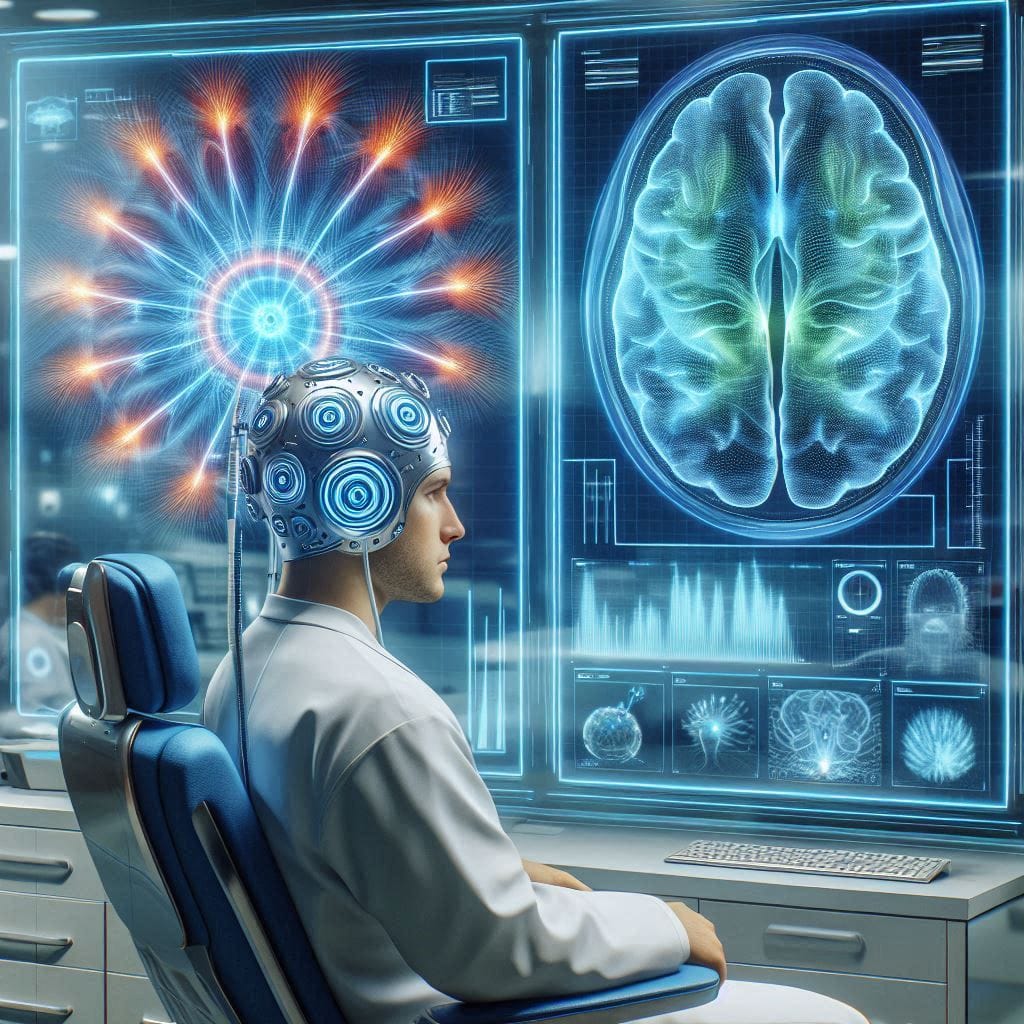
How Does a MEG Scan Work?
A MEG scan is a cutting-edge process that maps brain activity by detecting the magnetic fields produced by electrical currents in neurons. It is a completely non-invasive brain imaging procedure, ensuring both safety and comfort for the patient.
Equipment Details
The key to a MEG scan lies in its sophisticated hardware, particularly the SQUID sensors (Superconducting Quantum Interference Devices). These sensors, housed in a helmet-like device, are cooled to near absolute zero temperatures using liquid helium to achieve extreme sensitivity. The helmet is designed to fit snugly around the patient’s head to ensure accurate and comprehensive coverage of the brain’s magnetic activity.
The environment of the MEG room is also crucial. Since the magnetic fields generated by the brain are incredibly faint, the scan is performed in a magnetically shielded room to eliminate interference from external magnetic sources.
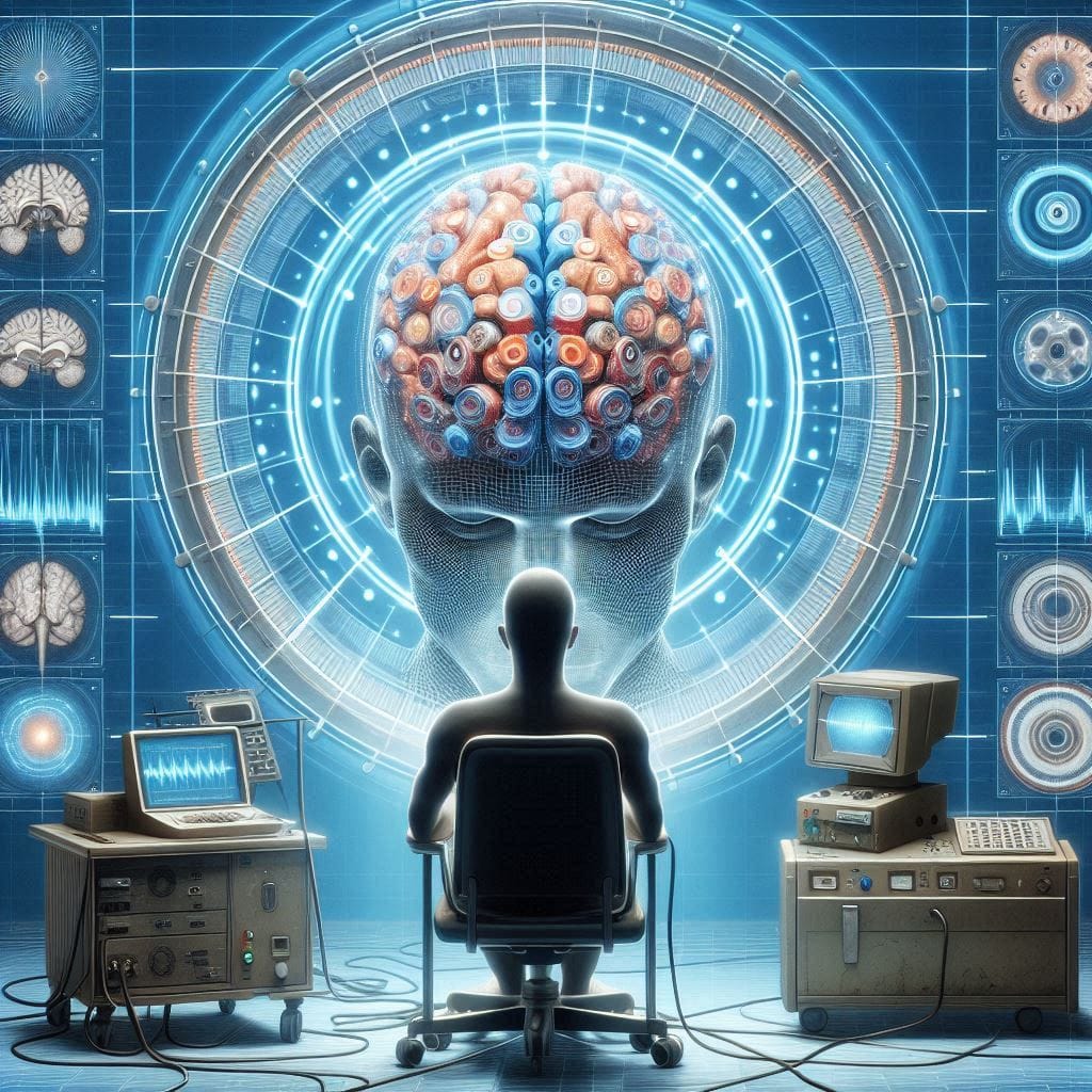
Step-by-Step Procedure
- Preparation
The patient is seated or reclined comfortably in the scanning chair. For children or individuals with anxiety, soothing techniques are often employed to ensure relaxation during the procedure. - Positioning
The helmet-like MEG device is positioned over the patient’s head. Markers may be placed on the scalp to help align the brain’s position relative to the scanner. - Data Collection
The patient is asked to remain still while performing specific tasks (e.g., moving a finger, viewing images, or listening to sounds) or while at rest. The MEG system records brain activity in real-time, capturing the magnetic fields produced by neuronal firing. - Duration
The scan typically lasts between 30 and 90 minutes, depending on the purpose of the test. Throughout, the patient can communicate with technicians to ensure comfort. - Post-Scan Analysis
Collected data is processed using advanced algorithms to create detailed maps of brain activity, which can then be compared to normative datasets.
Safety Measures and Patient Comfort
A MEG scan is entirely safe, as it does not involve exposure to radiation or invasive procedures. Patients can move freely after the test with no recovery time required. Its non-invasive nature makes it suitable for individuals of all ages, including children, and it is often favored over alternatives like invasive electrodes for brain mapping.
MEG vs. EEG
While EEG measures electrical activity, MEG captures the corresponding magnetic fields. This eliminates distortions caused by the scalp and skull, providing more precise spatial resolution. For deeper brain regions, MEG proves significantly more effective.
Applications of MEG Imaging
The versatility of Magnetoencephalography has made it an invaluable tool in both clinical and research settings. From diagnosing neurological disorders to uncovering the mysteries of cognition, the applications of MEG are transformative.
Clinical Uses
- Epilepsy
MEG is particularly effective in identifying the origins of epileptic seizures. By pinpointing seizure foci, it aids in presurgical planning to ensure that only affected areas are removed, preserving healthy brain tissue. - Brain Tumors
In cases of brain tumors, MEG assists in mapping areas responsible for vital functions like speech and motor activity. This information is critical for surgeons to avoid damaging functional areas during tumor removal. - Cognitive Disorders
MEG contributes to diagnosing and understanding conditions such as Alzheimer’s disease, autism spectrum disorders, and schizophrenia by mapping alterations in brain activity patterns.
Research Uses
- Studying Brain Networks
Researchers use functional brain imaging techniques like MEG to investigate how different brain regions communicate during various activities, such as problem-solving, language comprehension, and sensory perception. - Functional Connectivity
MEG helps visualize how neural pathways work together to process information, shedding light on the neural basis of cognition and behavior.
Role in Surgery Preparation
One of the most impactful applications of MEG is in presurgical planning. By precisely mapping the brain’s functional areas, MEG minimizes risks during surgeries for epilepsy, tumors, or other conditions. This ensures the preservation of critical regions, such as those controlling speech and motor functions.
Neurodevelopmental Disorders
MEG is particularly beneficial for children, as its non-invasive and radiation-free approach allows for safe brain imaging. It is used to study developmental disorders like autism, providing insights into how neural networks differ in affected individuals.
Advantages and Limitations of MEG
Magnetoencephalography (MEG) offers a range of benefits that make it a vital tool in neuroscience and clinical diagnostics. However, its challenges must also be addressed to fully understand its current and future potential.
Advantages of MEG
- High Temporal Resolution
MEG captures neural activity in milliseconds, making it one of the most temporally accurate imaging technologies available. This is crucial for studying dynamic processes such as:- Speech production and comprehension: MEG helps track the timing and flow of signals in language networks.
- Sensory processing: Researchers can map how stimuli like sound and light are processed in real time.
- Brain disorders: Conditions like epilepsy involve rapid neural activity, which MEG can pinpoint accurately.
- Non-Invasive Brain Imaging
Unlike some imaging techniques that expose patients to radiation (e.g., PET) or require invasive procedures (e.g., intracranial EEG), MEG is completely safe and painless. Its ability to work without altering the brain’s natural state makes it ideal for repeated use, especially in:- Children and vulnerable populations: It is often used in pediatric epilepsy and autism research.
- Longitudinal studies: MEG allows researchers to track changes over time without health risks.
- Real-Time Mapping
By measuring magnetic fields from active neurons, MEG provides immediate data about how brain regions interact. For example, it can:- Identify functional connectivity between regions.
- Assist in presurgical planning, determining which areas to avoid during brain surgery.
- Wide Applicability
MEG can adapt to diverse research needs, including studying healthy brains, neurological disorders, and developmental processes. Its precision and adaptability have led to breakthroughs in understanding complex conditions like schizophrenia and Alzheimer’s disease.
Limitations of MEG
- High Costs and Maintenance
The technology relies on superconducting quantum interference devices (SQUIDs) that operate at extremely low temperatures, requiring liquid helium. The financial burden includes:- Initial setup costs exceeding millions of dollars.
- Ongoing expenses for cooling systems and facility maintenance.
- Limited Global Accessibility
MEG imaging centers are concentrated in developed nations. For regions lacking funding or expertise, this limits opportunities for research and patient care. - Technological Constraints
- Magnetic shielding requirements: MEG systems must be housed in special facilities to block interference from external magnetic sources, such as urban electrical grids.
- Signal depth limitations: While MEG excels at detecting activity near the brain’s surface, deeper brain regions like the hippocampus are harder to study with this method.
- Complex Data Analysis
Interpreting MEG signals requires expertise in advanced computational models. As a result, smaller institutions may lack the resources to fully utilize the technology.
MEG vs. Other Brain Imaging Techniques
Comparing Magnetoencephalography (MEG) with other technologies highlights its unique strengths and situational uses.
MEG vs. EEG
- Signal Sensitivity and Accuracy
- EEG (Electroencephalography): Measures electrical signals from the scalp, which can be distorted by bone and tissue.
- MEG: Detects magnetic fields, which are unaffected by the skull, providing superior spatial resolution.
- Temporal Resolution
Both techniques excel in temporal accuracy, but MEG offers:- Precise localization of active brain regions.
- Clearer differentiation of simultaneous activities in separate areas.
- Use Cases
- EEG: More suitable for basic monitoring (e.g., sleep studies or seizure detection).
- MEG: Ideal for complex tasks like identifying functional connectivity or planning surgeries.
MEG vs. fMRI (Functional Magnetic Resonance Imaging)
- Temporal vs. Spatial Resolution
- fMRI: Offers high spatial resolution, ideal for mapping anatomical features and blood flow.
- MEG: Superior for time-sensitive studies, such as auditory and motor responses.
- Neural Basis of Signals
- fMRI: Measures blood-oxygen-level-dependent signals, which are indirect indicators of brain activity.
- MEG: Directly captures neuronal magnetic fields, providing more accurate insights into active neural pathways.
- Comfort and Safety
MEG is quieter and less confining than fMRI, improving patient comfort, especially for individuals with claustrophobia or sensory sensitivity.
MEG vs. PET (Positron Emission Tomography)
- Invasiveness
- PET: Requires radioactive tracers, posing risks, especially for repeated use.
- MEG: Completely non-invasive, making it safer for vulnerable groups.
- Use Scenarios
- PET: Effective for studying metabolic changes and neurochemical processes, such as in Alzheimer’s disease.
- MEG: Preferred for studying rapid neural interactions.
Practical Applications of MEG in Comparison
| Technology | Best Uses | Strengths |
|---|---|---|
| MEG | Functional mapping, epilepsy, research | High temporal resolution, non-invasive |
| EEG | Sleep studies, seizure detection | Affordable, portable |
| fMRI | Structural mapping, blood flow studies | High spatial resolution |
| PET | Metabolic studies, neurochemistry | Tracks biochemical activity |
Innovations and Future Prospects
Magnetoencephalography (MEG) is not only a cornerstone in current neuroscience and clinical diagnostics but also a rapidly evolving technology with exciting innovations and future applications. These advancements aim to overcome existing limitations and unlock new potential for MEG in both research and medical fields.
Wearable MEG Devices: Mobility and Ease of Use
Recent developments in wearable MEG systems are transforming the traditional static, room-confined setups.
- Technology Overview
- Wearable MEG devices utilize lightweight sensors, such as optically pumped magnetometers (OPMs), which eliminate the need for liquid helium cooling.
- Compact designs enable natural movements, such as walking or speaking, allowing real-world brain activity studies.
- Applications
- Children and Special Needs Populations: Wearable MEG provides comfort and better compliance in pediatric and neurodevelopmental studies.
- Sports and Rehabilitation: Athletes’ brain activities can now be monitored during motion-intensive tasks, aiding concussion studies and recovery planning.
- Challenges
- Ensuring sufficient sensitivity while maintaining portability.
- Developing cost-effective systems for widespread adoption.
AI and Machine Learning Integration
- Enhanced Data Analysis
MEG produces vast amounts of complex data, which can be difficult to interpret manually. Artificial intelligence (AI) and machine learning (ML) algorithms are addressing this by:- Automatically detecting patterns of neural connectivity.
- Classifying brain disorders with greater accuracy and speed, including early detection of Alzheimer’s or epilepsy.
- Predictive Modeling
- AI tools are being used to predict future brain activity based on current patterns, enabling preventive care and tailored interventions.
- Real-Time Insights
- ML algorithms are helping transform MEG from a diagnostic tool into a predictive and therapeutic system by offering real-time insights during surgeries or rehabilitation.
Broader Accessibility
- Technological Advancements
- Ongoing innovations aim to reduce the cost of MEG imaging systems, such as OPM sensors and advanced magnetic shielding technologies.
- Miniaturized and modular designs are under development to allow MEG systems in smaller clinics or mobile setups.
- Global Expansion
- Increasing awareness and funding in developing nations could bridge the accessibility gap, bringing MEG’s benefits to underserved regions.
Future Applications
- Brain-Computer Interfaces (BCIs)
MEG’s ability to capture precise brain signals could revolutionize BCIs, enabling more intuitive prosthetics and communication tools for people with disabilities. - Mental Health Diagnostics
- Detailed brain mapping could help diagnose and treat mental health conditions like depression, anxiety, and PTSD.
- Educational Tools
- Real-time brain monitoring may help in customizing learning approaches for students with varying cognitive abilities.
Conclusion
Magnetoencephalography (MEG) has firmly established itself as a transformative tool in neuroscience and medicine. From its unparalleled temporal resolution to its non-invasive nature, MEG has advanced our understanding of the brain and continues to improve patient outcomes.
Key Takeaways
- Revolutionizing Neuroscience
MEG enables real-time mapping of brain functions, opening new avenues for understanding and treating complex neurological conditions like epilepsy and Alzheimer’s disease. - A Tool for Precision Medicine
- Its role in presurgical planning and individualized treatment approaches highlights its importance in modern medicine.
Advancing Technology
-
- Wearable MEG devices and AI-driven analytics are paving the way for broader and more effective applications.
- With ongoing innovation, MEG is poised to become more accessible and affordable worldwide.
For researchers, clinicians, and students, understanding MEG’s potential is vital to leveraging its capabilities in neuroscience and beyond. Patients and caregivers should consult medical professionals to learn about MEG’s applications for specific conditions.
As advancements continue to address current limitations, Magnetoencephalography is set to redefine the possibilities in brain imaging, offering a brighter future for diagnostics, treatment, and research.
Dive Deeper into Brain and Technology Innovations
Expand your understanding of cutting-edge brain-related technologies and their transformative potential:
- Magnetogenetics and Beyond: Explore how genetic manipulation combined with magnetic fields is opening new frontiers in neuroscience and treatment modalities.
- The Evolution and Diagnostic Imaging History: Discover the fascinating journey of diagnostic imaging, from its origins to the advanced tools shaping modern medicine.
- Exploring Neuromorphic Computing: Learn how computers inspired by the human brain are revolutionizing artificial intelligence and machine learning.
- Exploring Brain-Computer Interface: Delve into the world of brain-computer interfaces, where technology bridges the gap between human thoughts and machine control.
These articles provide a broader perspective on the innovations that complement and enhance the understanding of brain imaging and neurotechnology.