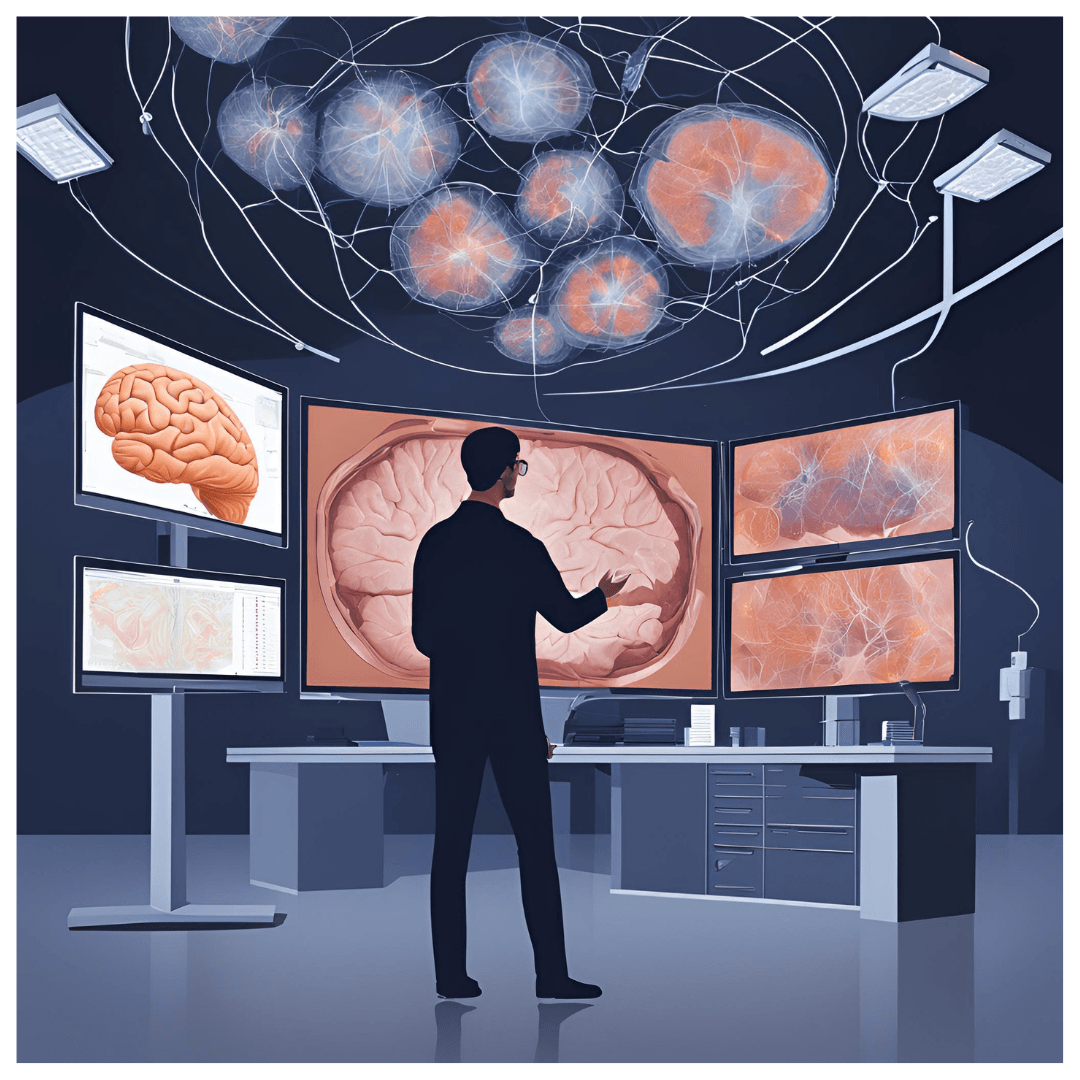
Introduction: The Importance of Neuroimaging in Modern Science
Neuroimaging has become a cornerstone of modern science, offering transformative insights into the human brain’s structure and function. By leveraging advanced technologies like MRI, CT scans, and PET scans, researchers and clinicians can visualize the intricate workings of the brain, diagnose neurological disorders, and unravel mysteries of cognition and behavior. These techniques not only enhance our understanding of complex neural networks but also drive innovations in medicine, psychology, and artificial intelligence. The evolution of neuroimaging continues to shape our approach to studying the brain and addressing global health challenges.
What Is Neuroimaging?
Neuroimaging is a collection of advanced, non-invasive technologies designed to visualize and analyze the structure and function of the brain. These techniques have been instrumental in revolutionizing neuroscience, enabling researchers to examine the brain in both health and disease without requiring surgical interventions.
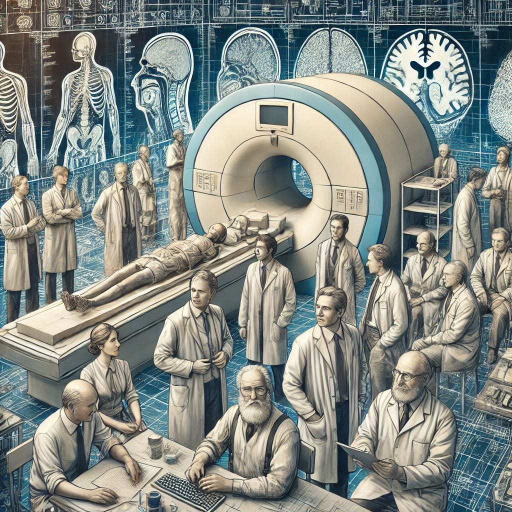
The field of neuroimaging began with the first recording of an electroencephalogram (EEG) by Hans Berger in 1924, marking the dawn of brain wave analysis. Over the decades, imaging technology advanced significantly, with the development of Computed Tomography (CT) in the 1970s and Magnetic Resonance Imaging (MRI) in the 1980s. Functional imaging, such as functional MRI (fMRI) and Positron Emission Tomography (PET), emerged later, providing insights into real-time brain activity.
Why Is Neuroimaging Important?
- Medical Applications: Neuroimaging plays a crucial role in diagnosing and managing conditions such as stroke, Alzheimer’s disease, epilepsy, and brain tumors. For instance, an MRI scan can detect structural anomalies, while a PET scan can reveal abnormal brain metabolism.
- Cognitive and Behavioral Research: Functional neuroimaging, like fMRI, has expanded our understanding of cognitive processes such as learning, memory, and decision-making.
- Non-Invasive and Safe: Unlike earlier invasive techniques, modern neuroimaging methods are safe for repeated use, enabling long-term studies and better monitoring of disease progression.
Impact on Neuroscience and Beyond
Neuroimaging has bridged the gap between behavioral psychology and biological neuroscience, allowing for a comprehensive understanding of brain-behavior relationships. For example, studies using fMRI have identified brain regions associated with empathy, creativity, and resilience.
“Advancements in neuroimaging techniques have made it possible to explore the intricacies of the human brain, leading to breakthroughs in medical diagnosis and cognitive research. By understanding what is neuroimaging techniques, we can appreciate their role in shaping modern neuroscience and healthcare.”
What Are Neuroimaging Techniques?
Neuroimaging techniques are scientific methods used to create detailed images of the brain’s structure and function. These methods fall into two main categories: structural neuroimaging and functional neuroimaging.
Structural Neuroimaging Techniques
Structural methods focus on visualizing the brain’s anatomy, providing insights into its physical structure and detecting abnormalities. Key techniques include:
- Magnetic Resonance Imaging (MRI): Uses magnetic fields and radio waves to generate high-resolution images of soft tissues. MRI is pivotal in diagnosing conditions like brain tumors, multiple sclerosis, and traumatic brain injury.
- Computed Tomography (CT): Combines X-rays to create cross-sectional images of the brain, useful for identifying acute conditions like hemorrhages or fractures.
Functional Neuroimaging Techniques
Functional methods measure dynamic processes within the brain, such as neural activity or blood flow. Key techniques include:
- Functional Magnetic Resonance Imaging (fMRI): Tracks changes in blood oxygen levels, mapping active brain regions during specific tasks. It is widely used in cognitive neuroscience.
- Positron Emission Tomography (PET): Utilizes radioactive tracers to monitor metabolic activity, often employed in research on dementia and cancer.
- Electroencephalography (EEG): Records electrical activity in the brain, critical for studying sleep disorders, epilepsy, and neural responses.
Applications of Neuroimaging Techniques
- Brain Mapping: Structural and functional imaging combined allows for detailed maps of the brain, highlighting areas responsible for specific functions.
- Disease Diagnosis: Early detection of neurological diseases, such as Parkinson’s or Alzheimer’s, is greatly aided by imaging techniques.
- Research and Development: Functional imaging facilitates studies on neuroplasticity, cognitive processes, and even the effects of drugs.
“Understanding what is neuroimaging techniques and their classification into structural and functional methods sheds light on how they revolutionize brain mapping and disease diagnosis. Modern neuroimaging techniques empower researchers to explore the brain’s complexities like never before.”
Structural Neuroimaging Techniques: Unlocking the Brain’s Anatomy
Structural neuroimaging techniques are pivotal tools in neuroscience and medicine, providing detailed images of the brain’s anatomy. They are crucial for diagnosing, studying, and monitoring various neurological conditions. This section delves into two primary methods: Computed Tomography (CT) and Magnetic Resonance Imaging (MRI), highlighting their use cases, advantages, and limitations.
Computed Tomography (CT) Scans
CT scans utilize X-ray technology to generate cross-sectional images of the brain. These scans are widely used for their speed and efficiency, especially in emergency situations.
Key Use Cases:
- Detecting brain injuries, such as hemorrhages or fractures.
- Identifying tumors, cysts, or structural abnormalities.
- Assessing stroke damage quickly.
Advantages:
- Speed: CT scans are quick, making them ideal for critical cases.
- Cost-Effective: Typically more affordable than MRI.
- Widespread Availability: Accessible in most healthcare settings.
Limitations:
- Radiation Exposure: Involves ionizing radiation, which limits repeated use.
- Lower Detail for Soft Tissue: Compared to MRI, CT provides less clarity for soft tissue structures like the brain’s gray and white matter.
Magnetic Resonance Imaging (MRI)
MRI uses powerful magnets and radio waves to produce highly detailed images of the brain’s structure without ionizing radiation.
Key Use Cases:
- Diagnosing multiple sclerosis, brain tumors, and vascular irregularities.
- Detecting subtle structural changes in neurodegenerative diseases like Alzheimer’s.
- Providing precise images for pre-surgical planning.
Advantages:
- Superior Detail: Especially effective for soft tissues and detecting minute abnormalities.
- No Radiation: Safer for repeated use.
- Customizable Imaging: Advanced techniques like T1-weighted and T2-weighted imaging allow for tailored diagnostics.
Limitations:
- Cost and Accessibility: More expensive and not as readily available as CT in some regions.
- Time-Consuming: Scans take longer, which can be challenging for emergency diagnostics.
- Claustrophobia Issues: The enclosed nature of the scanner may be uncomfortable for some patients.
Comparison: CT vs. MRI
| Feature | CT Scan | MRI Scan |
|---|---|---|
| Imaging Modality | X-rays | Magnetic fields and radio waves |
| Speed | Fast (5-10 minutes) | Longer (30-60 minutes) |
| Radiation Exposure | Yes | No |
| Detail Level | Moderate for soft tissue | High for soft tissue |
| Common Use Cases | Acute trauma, fractures | Chronic conditions, tumors |
When to Use Each Technique
- CT Scans are best for emergencies requiring immediate imaging, such as trauma or stroke.
- MRI Scans are the gold standard for detailed anatomical studies, particularly in chronic or complex conditions.
Visual Aid: Differences Between CT and MRI
To enhance understanding, include a side-by-side infographic:
- A CT image showing a brain hemorrhage.
- An MRI image highlighting a brain tumor. This visual comparison can help readers grasp the unique capabilities of each technique.
Structural neuroimaging techniques, including CT and MRI, are indispensable for visualizing the brain’s anatomy and addressing neurological challenges. While CT excels in emergencies, MRI offers unparalleled detail for in-depth analysis. Together, these tools continue to revolutionize brain science and patient care.
Functional Neuroimaging Techniques
Functional neuroimaging techniques focus on assessing the brain’s activity in real-time, providing insights into how different brain regions work together during cognitive tasks, emotions, and sensory processing. These techniques are crucial for understanding brain functions such as thought processes, learning, memory, and response to stimuli.
fMRI (Functional Magnetic Resonance Imaging)
How It Works: fMRI detects changes in blood oxygenation levels, which correlate with neural activity. When brain regions become active, they consume more oxygen, and fMRI can capture this dynamic shift in blood flow.
Key Use Cases:
- Cognitive Neuroscience: Studying processes like memory, decision-making, and language.
- Mental Disorders: Understanding brain activity patterns in conditions like depression, schizophrenia, and autism.
Advantages: High spatial resolution allows precise localization of brain activity. It is widely used in research to map brain functions.
Limitations: Lower temporal resolution compared to EEG. It’s also expensive and requires patients to remain still for extended periods.
PET (Positron Emission Tomography)
How It Works: PET scans track the movement of radioactive tracers injected into the bloodstream. These tracers are metabolized by brain cells, and PET detects this activity, which reflects neural function.
Key Use Cases:
- Brain Metabolism: PET is often used to assess metabolic activity in neurodegenerative diseases like Alzheimer’s and Parkinson’s.
- Tumor Detection: Used to identify abnormal metabolic patterns, such as cancerous growths in the brain.
Advantages: Provides functional information on metabolic activity, making it ideal for disease monitoring.
Limitations: Involves radioactive tracers, so repeated scans are not ideal. It’s also expensive and less widely available.
EEG (Electroencephalography)
How It Works: EEG measures the electrical activity of the brain by placing electrodes on the scalp to detect voltage fluctuations from neural oscillations.
Key Use Cases:
- Epilepsy: EEG is crucial in diagnosing and monitoring seizures.
- Sleep Disorders: It helps in studying the different stages of sleep and sleep disorders.
Advantages: High temporal resolution, making it perfect for tracking rapid changes in brain activity.
Limitations: Poor spatial resolution, limiting its ability to pinpoint the precise location of brain activity.
MEG (Magnetoencephalography)
How It Works: MEG detects the magnetic fields produced by neuronal activity, allowing for the measurement of brain activity with high temporal and spatial precision.
Key Use Cases:
- Cognitive Research: Understanding how the brain processes stimuli and complex functions like language.
- Epilepsy Monitoring: MEG can localize the source of epileptic seizures with high accuracy.
Advantages: Combines high temporal and spatial resolution, making it ideal for mapping brain functions.
Limitations: Expensive and requires specialized equipment.
fNIRS (Functional Near-Infrared Spectroscopy)
How It Works: fNIRS uses infrared light to detect changes in oxygenated and deoxygenated hemoglobin levels, providing insight into brain activity.
Key Use Cases:
- Cognitive Function: fNIRS is increasingly used to monitor cognitive activity in real-world settings, especially in children and the elderly.
- Neurorehabilitation: Useful for tracking brain activity in patients undergoing rehabilitation after strokes.
Advantages: Portable and cost-effective compared to other methods.
Limitations: Limited penetration depth, which makes it less effective for studying deeper brain structures.
Comparison Table of Functional Neuroimaging Techniques
| Technique | Temporal Resolution | Spatial Resolution | Cost | Common Use Cases |
|---|---|---|---|---|
| fMRI | Moderate | High | Expensive | Cognitive neuroscience, mental health disorders |
| PET | Low | Moderate | Very Expensive | Brain metabolism, cancer diagnosis |
| EEG | High | Low | Moderate | Epilepsy, sleep disorders |
| MEG | High | High | Very Expensive | Cognitive research, epilepsy |
| fNIRS | Moderate | Moderate | Low | Neurorehabilitation, real-world cognition |
“Functional neuroimaging techniques like fMRI, EEG, and PET are crucial tools in exploring brain activity and understanding complex brain functions. These methods help to monitor cognitive functions, identify neural abnormalities, and advance mental health research.”
Structural and Functional Neuroimaging Techniques: Bridging the Gap
Combining structural and functional neuroimaging techniques offers a comprehensive view of brain anatomy and activity. This integration provides a deeper understanding of brain structure-function relationships, enabling researchers to identify how specific regions contribute to cognitive processes and neurological disorders. The blending of these techniques is essential for mapping the brain in more detail and precision than either technique can alone.
fMRI and Structural MRI: A Powerful Combination
One of the most common hybrid approaches is the integration of functional MRI (fMRI) with structural MRI.
How It Works: Structural MRI provides high-resolution anatomical images of the brain, while fMRI tracks dynamic changes in brain activity. Combining these allows researchers to map which specific brain regions are involved in particular tasks and how those areas interact with the rest of the brain in real time.
Key Applications:
- Cognitive Mapping: This hybrid method allows scientists to not only locate brain activity but also see which anatomical structures are associated with cognitive functions such as memory, speech, and decision-making.
- Clinical Use: In patients with neurological disorders, combining structural and functional images helps pinpoint both damaged areas and alterations in brain activity, improving diagnosis and treatment planning.
Case Studies and Research Examples
- Epilepsy and Brain Surgery: Research combining fMRI and structural MRI has been instrumental in pre-surgical mapping for epilepsy patients. By identifying the precise areas of the brain where seizures originate, and understanding the surrounding structures, surgeons can plan safer, more targeted surgeries to remove epileptic foci while preserving essential brain functions.
- Parkinson’s Disease: Combining functional and structural neuroimaging techniques helps track both the physical degeneration of brain structures (like the substantia nigra) and the functional impairments in motor skills, improving treatment outcomes.
- Cognitive Neuroscience: Hybrid approaches are also used to understand how structural abnormalities (such as cortical thinning) correlate with functional deficits in mental health disorders like schizophrenia and dementia.
Future Trends in Hybrid Neuroimaging
The future of hybrid neuroimaging is promising, as newer technologies continue to push the boundaries of what’s possible:
- Improved Temporal and Spatial Resolution: Advancements in scanner technology are leading to better spatial and temporal resolution, allowing for more accurate and detailed brain mapping.
- Integration with AI and Machine Learning: AI-driven analysis of hybrid datasets promises to uncover hidden patterns in complex brain activity and structure, offering more personalized approaches to treatment.
- Multimodal Imaging: Researchers are increasingly combining techniques like fMRI, PET, EEG, and MEG to obtain both high temporal and spatial resolution for more comprehensive insights into brain function and structure.
Structural and functional neuroimaging techniques have profoundly transformed our understanding of the brain, offering unprecedented insights into its anatomy and activity. Combining methods like fMRI with structural MRI allows scientists and clinicians to explore both the brain’s anatomy and its activity in real time, enhancing both research and patient care.”
New and Emerging Neuroimaging Techniques
The field of neuroimaging is constantly evolving, with new advancements improving our ability to observe and understand the brain. Emerging neuroimaging techniques are breaking through existing limitations like resolution boundaries, cost, and the need for complex, invasive procedures. These developments open doors for more precise, real-time brain imaging and greater accessibility in clinical settings.
7T MRI (7 Tesla MRI)
How It Works: 7T MRI refers to the use of ultra-high magnetic field strength (7 Tesla), allowing for greater resolution compared to traditional MRI scanners (usually 1.5T or 3T).
Key Advantages:
- High-Resolution Imaging: This technique provides unprecedented anatomical detail, allowing for the visualization of very small structures within the brain, such as fine cortical layers or microvascular abnormalities.
- Applications: Researchers are using 7T MRI to study complex brain structures and disorders like multiple sclerosis, Alzheimer’s, and epilepsy. It’s particularly useful in areas where fine brain detail is needed for both structural and functional imaging.
Limitations: High cost, limited availability, and the need for specialized training.
AI-Integrated Imaging
How It Works: Artificial intelligence (AI) is being integrated into neuroimaging techniques, including MRI, PET, and CT. AI algorithms can analyze large datasets quickly and accurately, identifying patterns that might be missed by the human eye.

Key Advantages:
- Enhanced Accuracy: AI-enhanced imaging can reduce human error and increase diagnostic precision by providing automated analyses of scans.
- Real-Time Processing: AI can streamline real-time neuroimaging during surgical procedures or critical care, improving decision-making and patient outcomes.
Applications: AI is being used to help identify early signs of neurodegenerative diseases, as well as in planning surgeries for brain tumor removal and deep brain stimulation.
Multi-Modal Imaging Systems
How It Works: Multi-modal imaging combines data from multiple neuroimaging methods, such as fMRI, PET, and EEG, to capture both the anatomical and functional aspects of the brain at once.
Key Advantages:
- Comprehensive Insights: By combining the strengths of different techniques, multi-modal systems offer a fuller picture of brain activity, structure, and chemistry. For instance, fMRI and PET can be used together to study both brain metabolism and activity patterns simultaneously.
- Clinical Use: These systems are especially useful in personalized medicine, where a multi-faceted approach can help clinicians design more targeted treatment plans.
Limitations: Multi-modal imaging requires significant computational power and specialized equipment, which can make it costly and complex to implement in routine clinical practice.
Overcoming Existing Challenges
Emerging neuroimaging techniques are tackling several key challenges in the field:
- Resolution Limitations: New technologies like 7T MRI and multi-modal imaging offer higher resolution, helping to identify small and subtle changes in brain structures and activity that were previously difficult to detect.
- Cost Barriers: Though these techniques are initially expensive, advancements in AI and more efficient imaging algorithms may lower costs over time, making high-resolution imaging more accessible in the future.
- Invasive Nature: With developments in non-invasive neuroimaging methods, researchers can now observe the brain’s structure and function in real time, without the need for invasive procedures or radiation exposure, improving patient safety.
“New neuroimaging techniques such as 7T MRI, AI-integrated imaging, and multi-modal systems are revolutionizing the study of the brain, overcoming previous limitations in resolution, cost, and accessibility. These cutting-edge advancements are enabling more precise and real-time brain imaging, which is critical for both research and clinical applications.”
Neuroimaging in Psychology: Applications and Insights
Neuroimaging has become a vital tool in psychology, providing a window into the brain’s activity to better understand and diagnose various psychological conditions. Techniques like fMRI, PET, and EEG help to identify the brain’s neural mechanisms underlying mental health disorders, allowing for more accurate diagnoses and treatments. These tools give scientists the ability to map brain activity, which can offer insight into the root causes of psychological conditions, such as depression, anxiety, PTSD, and schizophrenia.
Applications in Mental Health Disorders
Depression:
- fMRI has been pivotal in understanding the neural correlates of depression. Studies have shown that depression is linked to abnormal brain activity in the prefrontal cortex and limbic system, areas responsible for regulating emotions. Research using fMRI has identified decreased activity in the prefrontal cortex and heightened activity in the amygdala in depressed individuals.
- Example Study: A study by Drevets et al. (2008) demonstrated abnormal activity in the subgenual cingulate cortex of depressed patients, further cementing the notion that specific brain regions are associated with depressive symptoms.
Post-Traumatic Stress Disorder (PTSD):
Neuroimaging has provided valuable insights into how trauma affects the brain. PTSD often correlates with hyperactivity in the amygdala (responsible for fear responses) and hypoactivity in the prefrontal cortex (involved in emotion regulation). fMRI has helped establish these links, making it easier to diagnose and treat PTSD.
- Example Study: Felmingham et al. (2014) showed that individuals with PTSD had heightened amygdala activity when exposed to trauma-related stimuli, providing crucial information for targeted therapies.
Schizophrenia:
Schizophrenia is characterized by abnormalities in brain structure and function. Structural and functional neuroimaging, including fMRI and PET, has revealed altered connectivity between brain regions such as the frontal cortex, hippocampus, and thalamus in patients with schizophrenia.
- Example Study: Research by Stephan et al. (2006) using PET imaging identified reduced dopamine function in the prefrontal cortex of schizophrenia patients, which is a critical finding in understanding the pathophysiology of the disorder.
fMRI Applications in Anxiety Research
Functional MRI (fMRI) has been widely used to study anxiety. Studies reveal that anxiety disorders are often linked to overactivity in the amygdala and underactivity in the prefrontal cortex. fMRI helps to visualize these patterns and is increasingly used to assess treatments, such as cognitive behavioral therapy (CBT) or pharmacological interventions.
- Example Study: Research by Hughes et al. (2009) demonstrated increased amygdala activity in individuals with social anxiety disorder when exposed to anxiety-provoking stimuli, highlighting the potential of fMRI in tailoring specific treatments.
The integration of neuroimaging techniques in psychological research provides an invaluable tool for understanding mental health disorders at a neural level. By pinpointing the areas of the brain involved in conditions like depression, PTSD, and schizophrenia, neuroimaging is paving the way for more accurate diagnoses, targeted therapies, and better patient outcomes.
Ethical Considerations and Challenges in Neuroimaging
While neuroimaging techniques have opened new frontiers in medical and psychological research, they also raise significant ethical concerns. As brain imaging becomes more advanced, the questions of data privacy, informed consent, and the potential misuse of neuroimaging data must be carefully addressed.
- Data Privacy and Security:
Neuroimaging studies involve sensitive personal data about brain activity and mental health. If this data were to be hacked or misused, it could lead to privacy violations or discrimination. Establishing strict data protection protocols is vital to mitigate these risks. - Informed Consent:
One of the major ethical challenges in neuroimaging is obtaining informed consent, particularly in vulnerable populations. Participants must fully understand the nature of the study, the potential risks involved, and the implications of their brain data being used for research. Clear communication is essential to ensure that participants’ rights are respected. - Neuroethics of Brain Manipulation:
As neuroimaging technologies advance, they may be used in conjunction with interventions that alter brain activity, such as deep brain stimulation or neuromodulation. The potential for such interventions to change aspects of cognition, memory, and personality raises ethical concerns about autonomy and identity.
Technical Challenges in Neuroimaging
While the advancements in neuroimaging are groundbreaking, several technical challenges remain:
- Cost and Accessibility:
High-end neuroimaging equipment, like fMRI, PET, and 7T MRI, are expensive, limiting their availability to only well-funded institutions or research centers. This creates barriers for many researchers and healthcare providers, particularly in low-income regions. - Training Requirements:
Neuroimaging techniques require highly specialized training to operate and interpret correctly. Without proper expertise, there’s a risk of misinterpretation of results, which could lead to inaccurate diagnoses or treatments. - Resolution and Speed:
While newer technologies are improving resolution and speed, there is still a trade-off between image clarity and the time required to obtain these images. High-resolution scans can take much longer, which might not always be practical in clinical settings.
The continued advancement of neuroimaging technologies raises both opportunities and challenges. While they promise significant breakthroughs in understanding the brain, ethical and technical concerns must be navigated carefully. Ensuring privacy, accessibility, and proper training will be essential for maximizing the benefits of these powerful tools.
Conclusion: The Future of Neuroimaging
Transformative Potential
Neuroimaging has already revolutionized our understanding of the brain, but its full potential is yet to be realized. The combination of structural and functional imaging techniques has provided unprecedented insights into both brain anatomy and activity. As technologies like 7T MRI, AI-integrated imaging, and multi-modal systems evolve, the ability to visualize and understand the brain will continue to improve, opening new avenues for diagnosing and treating neurological and psychological conditions.
Call to Action
As we look to the future, the continued development of neuroimaging techniques will undoubtedly reshape the landscape of neuroscience, psychology, and medicine. To stay informed about the latest advancements in neuroimaging and its applications, explore our blog and share this post with others interested in the cutting-edge of brain research.
For a more comprehensive look, check out these related articles: